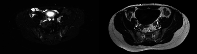
Prevalence of sacroiliitis on magnetic resonance enterography in Crohn's disease and its association with intestinal findings: a monocentric observational cross-sectional study.
G Amati, G Sandri, A Bertani, D Vaccari, A Pecchi, B Bongiovanni, M Orlandi, G Ciancio, M Pecchini, O Secchi, A Colecchia, P Torricelli, D Giuggioli
{"title":"Prevalence of sacroiliitis on magnetic resonance enterography in Crohn's disease and its association with intestinal findings: a monocentric observational cross-sectional study.","authors":"G Amati, G Sandri, A Bertani, D Vaccari, A Pecchi, B Bongiovanni, M Orlandi, G Ciancio, M Pecchini, O Secchi, A Colecchia, P Torricelli, D Giuggioli","doi":"10.1007/s10238-025-01846-1","DOIUrl":null,"url":null,"abstract":"<p><p>Magnetic resonance enterography (MRE) is recommended for the assessment of small intestine alterations in Crohn's disease (CD). Sacroiliac joints (SIJs) imaging has a central role in the early diagnosis of sacroiliitis (SI). MRE can evaluate both acute and structural findings of SIJs. We aimed to assess the prevalence of SI detected by MRE in a cohort of CD patients, and the associations of SI with demographic and clinical features and with intestinal MRE findings. Two hundred patients affected by CD (M:F 1:1, median age 49.5 (22.5) years, median CD duration 4.75 (16.2) years) tested with MRE between 2011 and 2023 were selected. They discontinued tumor necrosis factor α inhibitors (TNFαi) at least 3 months before the MRE execution. Most patients had an ileal CD location (65.0%) and a stricturing behavior of disease (50.0%). Thirty-five percent of patients underwent ileocecal resection. One out of ten patients were treated with at least one TNFαi. Active SI, capsulitis, erosions, sclerosis, and ankylosis were present in 10.5%, 0.5%, 2.0%, 2.5%, and 1.5%, respectively. No significant correlations have been evidenced between the presence of SI and demographic and clinical variables. The presence of an asymmetric hyperenhancement of the bowel wall was instead directly associated with the presence of SI (OR 8.61, 95% CI 1.47-50.4, p = 0.017). In this study, subclinical SI is a frequent finding in CD patients being present in one out of ten MRE examination. This phenomenon was significantly associated with asymmetric mural enhancement, a specific CD intestinal lesion at MRE.</p>","PeriodicalId":10337,"journal":{"name":"Clinical and Experimental Medicine","volume":"25 1","pages":"314"},"PeriodicalIF":3.5000,"publicationDate":"2025-09-04","publicationTypes":"Journal Article","fieldsOfStudy":null,"isOpenAccess":false,"openAccessPdf":"https://www.ncbi.nlm.nih.gov/pmc/articles/PMC12408789/pdf/","citationCount":"0","resultStr":null,"platform":"Semanticscholar","paperid":null,"PeriodicalName":"Clinical and Experimental Medicine","FirstCategoryId":"3","ListUrlMain":"https://doi.org/10.1007/s10238-025-01846-1","RegionNum":4,"RegionCategory":"医学","ArticlePicture":[],"TitleCN":null,"AbstractTextCN":null,"PMCID":null,"EPubDate":"","PubModel":"","JCR":"Q2","JCRName":"MEDICINE, RESEARCH & EXPERIMENTAL","Score":null,"Total":0}
引用次数: 0
Abstract
Magnetic resonance enterography (MRE) is recommended for the assessment of small intestine alterations in Crohn's disease (CD). Sacroiliac joints (SIJs) imaging has a central role in the early diagnosis of sacroiliitis (SI). MRE can evaluate both acute and structural findings of SIJs. We aimed to assess the prevalence of SI detected by MRE in a cohort of CD patients, and the associations of SI with demographic and clinical features and with intestinal MRE findings. Two hundred patients affected by CD (M:F 1:1, median age 49.5 (22.5) years, median CD duration 4.75 (16.2) years) tested with MRE between 2011 and 2023 were selected. They discontinued tumor necrosis factor α inhibitors (TNFαi) at least 3 months before the MRE execution. Most patients had an ileal CD location (65.0%) and a stricturing behavior of disease (50.0%). Thirty-five percent of patients underwent ileocecal resection. One out of ten patients were treated with at least one TNFαi. Active SI, capsulitis, erosions, sclerosis, and ankylosis were present in 10.5%, 0.5%, 2.0%, 2.5%, and 1.5%, respectively. No significant correlations have been evidenced between the presence of SI and demographic and clinical variables. The presence of an asymmetric hyperenhancement of the bowel wall was instead directly associated with the presence of SI (OR 8.61, 95% CI 1.47-50.4, p = 0.017). In this study, subclinical SI is a frequent finding in CD patients being present in one out of ten MRE examination. This phenomenon was significantly associated with asymmetric mural enhancement, a specific CD intestinal lesion at MRE.
期刊介绍:
Clinical and Experimental Medicine (CEM) is a multidisciplinary journal that aims to be a forum of scientific excellence and information exchange in relation to the basic and clinical features of the following fields: hematology, onco-hematology, oncology, virology, immunology, and rheumatology. The journal publishes reviews and editorials, experimental and preclinical studies, translational research, prospectively designed clinical trials, and epidemiological studies. Papers containing new clinical or experimental data that are likely to contribute to changes in clinical practice or the way in which a disease is thought about will be given priority due to their immediate importance. Case reports will be accepted on an exceptional basis only, and their submission is discouraged. The major criteria for publication are clarity, scientific soundness, and advances in knowledge. In compliance with the overwhelmingly prevailing request by the international scientific community, and with respect for eco-compatibility issues, CEM is now published exclusively online.
 求助内容:
求助内容: 应助结果提醒方式:


