{"title":"Electroacupuncture therapy improves cognitive dysfunction after ischemic stroke in Sprague-Dawley rats by adjusting the lncRNA-MEG3/miR-4640-3p axis.","authors":"Yun Zhang, Shiqing Gao, Ling Lin, Yongbing Zheng","doi":"10.1097/MS9.0000000000003628","DOIUrl":null,"url":null,"abstract":"<p><strong>Background: </strong>Ischemic stroke (IS) is a common disease that can cause cognitive dysfunction. Electroacupuncture (EA) is an effective way to alleviate cognitive dysfunction, but its molecular regulatory mechanism is still unclear. Long noncoding RNA-MEG3 (MEG3) is an important factor in the incidence and progression of IS. Herein, we explored the mechanism of EA in IS.</p><p><strong>Methods: </strong>A middle cerebral artery occlusion (MCAO) model was established in Sprague-Dawley rats to simulate IS <i>in vivo</i>, followed by electroacupuncture (EA) therapy. AAV-control and AAV-MEG3 were injected into the lateral ventricle of rats. All rats except for the sham group underwent MCAO. EA was performed at Shenting and Baihui points for 30 min, once a day for 14 days. The MEG3 and miR-4640-3p levels in brains were measured by qRT-PCR. Dual-luciferase reporter analysis validated the relationship between MEG3 and miR-4640-3p. The Morris water maze test and the neurological function test were carried out. The pathological morphology of the brain tissue was evaluated by H&E staining. Apoptotic cells in brains were examined utilizing TUNEL staining. The contents of Bax, Bcl-2, caspase-3, and CytC were assessed by western blot.</p><p><strong>Results: </strong>EA treatment reduced the content of MEG3 but enhanced miR-4640-3p levels in MCAO rats. MEG3 was a sponge for miR-4640-3p. EA treatment alleviated cognitive dysfunction in MCAO rats by inhibiting MEG3. EA treatment reduced MCAO-induced neural damage and apoptosis by inhibiting MEG3.</p><p><strong>Conclusion: </strong>EA improved cognitive dysfunction in IS rats by adjusting the MEG3/miR-4640-3p axis, suggesting that EA may be an effective potential therapeutic strategy for improvement of cognitive dysfunction in IS. This study provided a more reliable experimental basis for clinical EA treatment of IS patients.</p>","PeriodicalId":8025,"journal":{"name":"Annals of Medicine and Surgery","volume":"87 9","pages":"5512-5521"},"PeriodicalIF":1.6000,"publicationDate":"2025-07-25","publicationTypes":"Journal Article","fieldsOfStudy":null,"isOpenAccess":false,"openAccessPdf":"https://www.ncbi.nlm.nih.gov/pmc/articles/PMC12401418/pdf/","citationCount":"0","resultStr":null,"platform":"Semanticscholar","paperid":null,"PeriodicalName":"Annals of Medicine and Surgery","FirstCategoryId":"1085","ListUrlMain":"https://doi.org/10.1097/MS9.0000000000003628","RegionNum":0,"RegionCategory":null,"ArticlePicture":[],"TitleCN":null,"AbstractTextCN":null,"PMCID":null,"EPubDate":"2025/9/1 0:00:00","PubModel":"eCollection","JCR":"Q2","JCRName":"MEDICINE, GENERAL & INTERNAL","Score":null,"Total":0}
引用次数: 0
Abstract
Background: Ischemic stroke (IS) is a common disease that can cause cognitive dysfunction. Electroacupuncture (EA) is an effective way to alleviate cognitive dysfunction, but its molecular regulatory mechanism is still unclear. Long noncoding RNA-MEG3 (MEG3) is an important factor in the incidence and progression of IS. Herein, we explored the mechanism of EA in IS.
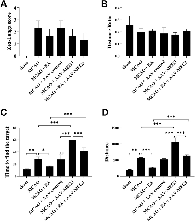
Methods: A middle cerebral artery occlusion (MCAO) model was established in Sprague-Dawley rats to simulate IS in vivo, followed by electroacupuncture (EA) therapy. AAV-control and AAV-MEG3 were injected into the lateral ventricle of rats. All rats except for the sham group underwent MCAO. EA was performed at Shenting and Baihui points for 30 min, once a day for 14 days. The MEG3 and miR-4640-3p levels in brains were measured by qRT-PCR. Dual-luciferase reporter analysis validated the relationship between MEG3 and miR-4640-3p. The Morris water maze test and the neurological function test were carried out. The pathological morphology of the brain tissue was evaluated by H&E staining. Apoptotic cells in brains were examined utilizing TUNEL staining. The contents of Bax, Bcl-2, caspase-3, and CytC were assessed by western blot.
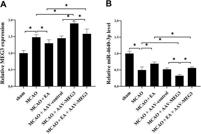
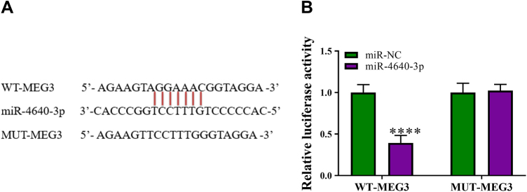
Results: EA treatment reduced the content of MEG3 but enhanced miR-4640-3p levels in MCAO rats. MEG3 was a sponge for miR-4640-3p. EA treatment alleviated cognitive dysfunction in MCAO rats by inhibiting MEG3. EA treatment reduced MCAO-induced neural damage and apoptosis by inhibiting MEG3.
Conclusion: EA improved cognitive dysfunction in IS rats by adjusting the MEG3/miR-4640-3p axis, suggesting that EA may be an effective potential therapeutic strategy for improvement of cognitive dysfunction in IS. This study provided a more reliable experimental basis for clinical EA treatment of IS patients.
 求助内容:
求助内容: 应助结果提醒方式:


