Junwei Dai, Jun Tao, Shusheng Wei, Baokun Han, Shuai Ma, Chengyu Wu, Long Sun, Xiaodong Ma, Yimeng Chen
{"title":"The mechanism of NK cell expression of RANKL/RANK/OPG pathway in mouse models of femoral head necrosis.","authors":"Junwei Dai, Jun Tao, Shusheng Wei, Baokun Han, Shuai Ma, Chengyu Wu, Long Sun, Xiaodong Ma, Yimeng Chen","doi":"10.1097/MS9.0000000000003615","DOIUrl":null,"url":null,"abstract":"<p><p>This study aimed to investigate the role of natural killer (NK) cells in the RANKL/RANK/OPG pathway in osteonecrosis of the femoral head (ONFH). C57 mice were categorized into a control group, an observation group (10 mice each), and an experimental group comprising 4 NK cell knockout mice. A hormone-induced femoral head necrosis model was created by administering lipopolysaccharide combined with methylprednisolone for 8 weeks to the experimental and control groups. The observation group received subcutaneous injections of an equal amount of normal saline. After 8 weeks, peripheral blood was collected from the mice, and bilateral femoral head specimens were obtained post-mortem. Expression levels of NK cells, OPG, RANK, and RANKL in the peripheral blood and joint fluid of ONFH mice were determined using PCR and ELISA techniques, and compared with the control group. The experimental group exhibited an increased number of NK cells in the peripheral blood and joint fluid compared to the control group. OPG expression was downregulated, while RANK and RANKL were significantly upregulated, resulting in a marked increase in the number of mature osteoclasts. In ONFH patients, NK cells were found to upregulate TNF-α and RANKL, downregulate IFN-γ and OPG, promote osteoclast maturation, disrupt bone balance, accelerate femoral head necrosis collapse, and ultimately hasten the progression of femoral head necrosis.</p>","PeriodicalId":8025,"journal":{"name":"Annals of Medicine and Surgery","volume":"87 9","pages":"5494-5502"},"PeriodicalIF":1.6000,"publicationDate":"2025-07-18","publicationTypes":"Journal Article","fieldsOfStudy":null,"isOpenAccess":false,"openAccessPdf":"https://www.ncbi.nlm.nih.gov/pmc/articles/PMC12401202/pdf/","citationCount":"0","resultStr":null,"platform":"Semanticscholar","paperid":null,"PeriodicalName":"Annals of Medicine and Surgery","FirstCategoryId":"1085","ListUrlMain":"https://doi.org/10.1097/MS9.0000000000003615","RegionNum":0,"RegionCategory":null,"ArticlePicture":[],"TitleCN":null,"AbstractTextCN":null,"PMCID":null,"EPubDate":"2025/9/1 0:00:00","PubModel":"eCollection","JCR":"Q2","JCRName":"MEDICINE, GENERAL & INTERNAL","Score":null,"Total":0}
引用次数: 0
Abstract
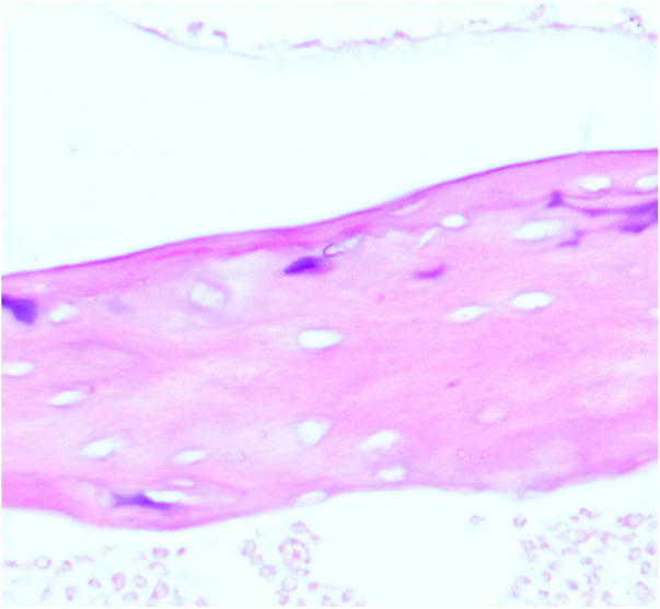
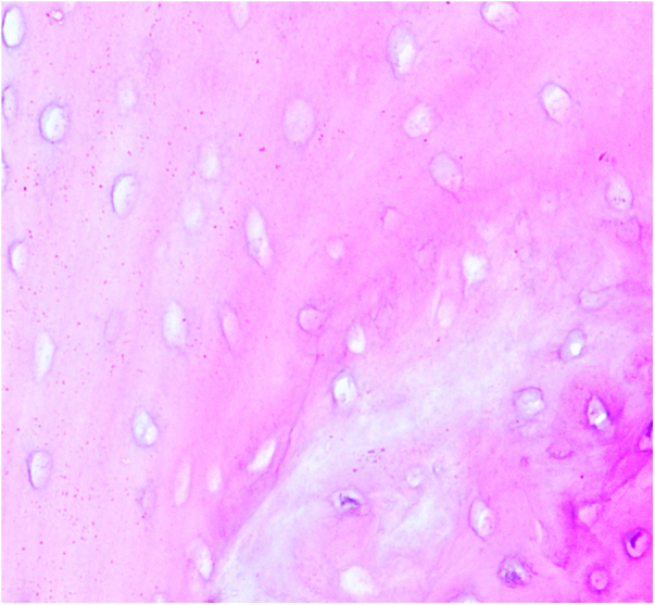
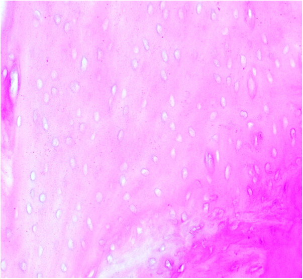
This study aimed to investigate the role of natural killer (NK) cells in the RANKL/RANK/OPG pathway in osteonecrosis of the femoral head (ONFH). C57 mice were categorized into a control group, an observation group (10 mice each), and an experimental group comprising 4 NK cell knockout mice. A hormone-induced femoral head necrosis model was created by administering lipopolysaccharide combined with methylprednisolone for 8 weeks to the experimental and control groups. The observation group received subcutaneous injections of an equal amount of normal saline. After 8 weeks, peripheral blood was collected from the mice, and bilateral femoral head specimens were obtained post-mortem. Expression levels of NK cells, OPG, RANK, and RANKL in the peripheral blood and joint fluid of ONFH mice were determined using PCR and ELISA techniques, and compared with the control group. The experimental group exhibited an increased number of NK cells in the peripheral blood and joint fluid compared to the control group. OPG expression was downregulated, while RANK and RANKL were significantly upregulated, resulting in a marked increase in the number of mature osteoclasts. In ONFH patients, NK cells were found to upregulate TNF-α and RANKL, downregulate IFN-γ and OPG, promote osteoclast maturation, disrupt bone balance, accelerate femoral head necrosis collapse, and ultimately hasten the progression of femoral head necrosis.
 求助内容:
求助内容: 应助结果提醒方式:


