Development of a methodology for the volume estimation of the prefrontal cortical subfields in very pre-term infants using magnetic resonance imaging and stereology.
{"title":"Development of a methodology for the volume estimation of the prefrontal cortical subfields in very pre-term infants using magnetic resonance imaging and stereology.","authors":"Faten Aldhafeeri","doi":"10.2478/abm-2025-0023","DOIUrl":null,"url":null,"abstract":"<p><strong>Background: </strong>The prefrontal cortex (PFC) is vital for cognitive and emotional functions and is vulnerable to disruptions in preterm infants. Reliable volume estimation methods are needed to study its development.</p><p><strong>Objective: </strong>To develop and validate a novel method for estimating the volume of PFC subfields in very preterm infants using magnetic resonance imaging (MRI) combined with stereological techniques. The method was designed to achieve a coefficient of error (CE) below 5%.</p><p><strong>Methods: </strong>Five preterm infants born before 28 weeks of gestation were scanned using a 1.5-Tesla MRI scanner. The points of intersection between the grid and structure boundaries, in addition to the points in each slice, were counted using in-house software (Easy Measure).</p><p><strong>Results: </strong>The shape coefficient for each subfield of the prefrontal cortex was calculated, which yielded coefficients of 4.5, 6.1, 6.4, and 6.5 for dorsolateral, dorsomedial, orbitolateral, and orbitomedial PFC regions, respectively. For the dorsolateral prefrontal cortex, a grid size of 4 × 4 pixels and a 0.2 cm slice gap for the dorsomedial prefrontal cortex (DMPFC), a grid size of 5 × 5 pixels and a 0.1 cm slice gap for the orbitolateral PFC, a grid size of 5 × 5 pixels and a 0.3 cm slice gap, and a grid size of 5 × 5 pixels and 0.1 cm slice gap for the DMPFC resulted in <5% CE.</p><p><strong>Conclusion: </strong>This methodology offers new insights into the neurodevelopmental effects of preterm birth and has potential applications in the early detection of neurodevelopmental disorders. Its precision, reliability, and non-invasive nature make it suitable for longitudinal studies and contribute to neonatal neuroimaging and neurodevelopmental research.</p>","PeriodicalId":8501,"journal":{"name":"Asian Biomedicine","volume":"19 4","pages":"174-182"},"PeriodicalIF":0.9000,"publicationDate":"2025-09-02","publicationTypes":"Journal Article","fieldsOfStudy":null,"isOpenAccess":false,"openAccessPdf":"https://www.ncbi.nlm.nih.gov/pmc/articles/PMC12404656/pdf/","citationCount":"0","resultStr":null,"platform":"Semanticscholar","paperid":null,"PeriodicalName":"Asian Biomedicine","FirstCategoryId":"3","ListUrlMain":"https://doi.org/10.2478/abm-2025-0023","RegionNum":4,"RegionCategory":"医学","ArticlePicture":[],"TitleCN":null,"AbstractTextCN":null,"PMCID":null,"EPubDate":"2025/8/1 0:00:00","PubModel":"eCollection","JCR":"Q4","JCRName":"MEDICINE, RESEARCH & EXPERIMENTAL","Score":null,"Total":0}
引用次数: 0
Abstract
Background: The prefrontal cortex (PFC) is vital for cognitive and emotional functions and is vulnerable to disruptions in preterm infants. Reliable volume estimation methods are needed to study its development.
Objective: To develop and validate a novel method for estimating the volume of PFC subfields in very preterm infants using magnetic resonance imaging (MRI) combined with stereological techniques. The method was designed to achieve a coefficient of error (CE) below 5%.
Methods: Five preterm infants born before 28 weeks of gestation were scanned using a 1.5-Tesla MRI scanner. The points of intersection between the grid and structure boundaries, in addition to the points in each slice, were counted using in-house software (Easy Measure).
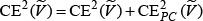
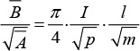
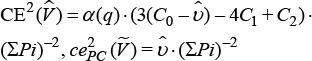
Results: The shape coefficient for each subfield of the prefrontal cortex was calculated, which yielded coefficients of 4.5, 6.1, 6.4, and 6.5 for dorsolateral, dorsomedial, orbitolateral, and orbitomedial PFC regions, respectively. For the dorsolateral prefrontal cortex, a grid size of 4 × 4 pixels and a 0.2 cm slice gap for the dorsomedial prefrontal cortex (DMPFC), a grid size of 5 × 5 pixels and a 0.1 cm slice gap for the orbitolateral PFC, a grid size of 5 × 5 pixels and a 0.3 cm slice gap, and a grid size of 5 × 5 pixels and 0.1 cm slice gap for the DMPFC resulted in <5% CE.
Conclusion: This methodology offers new insights into the neurodevelopmental effects of preterm birth and has potential applications in the early detection of neurodevelopmental disorders. Its precision, reliability, and non-invasive nature make it suitable for longitudinal studies and contribute to neonatal neuroimaging and neurodevelopmental research.
期刊介绍:
Asian Biomedicine: Research, Reviews and News (ISSN 1905-7415 print; 1875-855X online) is published in one volume (of 6 bimonthly issues) a year since 2007. [...]Asian Biomedicine is an international, general medical and biomedical journal that aims to publish original peer-reviewed contributions dealing with various topics in the biomedical and health sciences from basic experimental to clinical aspects. The work and authorship must be strongly affiliated with a country in Asia, or with specific importance and relevance to the Asian region. The Journal will publish reviews, original experimental studies, observational studies, technical and clinical (case) reports, practice guidelines, historical perspectives of Asian biomedicine, clinicopathological conferences, and commentaries
Asian biomedicine is intended for a broad and international audience, primarily those in the health professions including researchers, physician practitioners, basic medical scientists, dentists, educators, administrators, those in the assistive professions, such as nurses, and the many types of allied health professionals in research and health care delivery systems including those in training.
 求助内容:
求助内容: 应助结果提醒方式:


