R Praveen, Gianluca Scalia, Giuseppe Emmanuele Umana, Bipin Chaurasia
{"title":"Symptomatic postoperative cyst following glioma excision in a pediatric patient: a case report.","authors":"R Praveen, Gianluca Scalia, Giuseppe Emmanuele Umana, Bipin Chaurasia","doi":"10.1097/MS9.0000000000003666","DOIUrl":null,"url":null,"abstract":"<p><strong>Introduction and importance: </strong>Postoperative cyst formation is a rare but significant complication following central nervous system (CNS) tumor resection. This case report describes a 14-year-old girl who developed a postoperative cyst after glioma excision, successfully managed through a minimally invasive approach.</p><p><strong>Case presentation: </strong>A 14-year-old female presented with headache and seizures. Magnetic Resonance Imaging (MRI) revealed a mass in the trigonal region. She underwent surgical resection of the tumor. On postoperative day 7, she developed a symptomatic cyst at the tumor bed. The cyst was drained, resulting in symptomatic improvement. Follow-up assessments confirmed her clinical recovery.</p><p><strong>Clinical discussion: </strong>In pediatric patients, postoperative cyst formation is less frequently reported compared to adults. This highlights the need for further investigation into its underlying mechanisms and optimal treatment strategies in children.</p><p><strong>Conclusion: </strong>Minimally invasive percutaneous drainage followed by shunting successfully managed the postoperative cyst and prevented recurrence in this pediatric glioma case. Given the absence of standardized treatment protocols, individualized care is essential to ensure the best possible outcomes.</p>","PeriodicalId":8025,"journal":{"name":"Annals of Medicine and Surgery","volume":"87 9","pages":"6201-6205"},"PeriodicalIF":1.6000,"publicationDate":"2025-08-02","publicationTypes":"Journal Article","fieldsOfStudy":null,"isOpenAccess":false,"openAccessPdf":"https://www.ncbi.nlm.nih.gov/pmc/articles/PMC12401426/pdf/","citationCount":"0","resultStr":null,"platform":"Semanticscholar","paperid":null,"PeriodicalName":"Annals of Medicine and Surgery","FirstCategoryId":"1085","ListUrlMain":"https://doi.org/10.1097/MS9.0000000000003666","RegionNum":0,"RegionCategory":null,"ArticlePicture":[],"TitleCN":null,"AbstractTextCN":null,"PMCID":null,"EPubDate":"2025/9/1 0:00:00","PubModel":"eCollection","JCR":"Q2","JCRName":"MEDICINE, GENERAL & INTERNAL","Score":null,"Total":0}
引用次数: 0
Abstract
Introduction and importance: Postoperative cyst formation is a rare but significant complication following central nervous system (CNS) tumor resection. This case report describes a 14-year-old girl who developed a postoperative cyst after glioma excision, successfully managed through a minimally invasive approach.
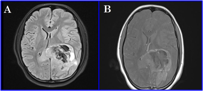
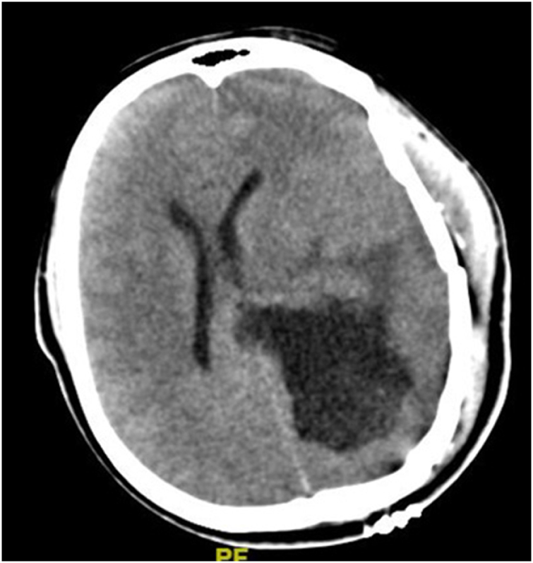
Case presentation: A 14-year-old female presented with headache and seizures. Magnetic Resonance Imaging (MRI) revealed a mass in the trigonal region. She underwent surgical resection of the tumor. On postoperative day 7, she developed a symptomatic cyst at the tumor bed. The cyst was drained, resulting in symptomatic improvement. Follow-up assessments confirmed her clinical recovery.
Clinical discussion: In pediatric patients, postoperative cyst formation is less frequently reported compared to adults. This highlights the need for further investigation into its underlying mechanisms and optimal treatment strategies in children.
Conclusion: Minimally invasive percutaneous drainage followed by shunting successfully managed the postoperative cyst and prevented recurrence in this pediatric glioma case. Given the absence of standardized treatment protocols, individualized care is essential to ensure the best possible outcomes.
 求助内容:
求助内容: 应助结果提醒方式:


