Haider Sarwar, Nicholas Tan, Saif Aldeen Alryalat, Cara Elise Capitena Young, Leonard K Seibold, Malik Y Kahook
{"title":"Acute Structural Effects of Novel Endoscopic Cyclophotocoagulation versus Standard Endoscopic and Transscleral Cyclophotocoagulation.","authors":"Haider Sarwar, Nicholas Tan, Saif Aldeen Alryalat, Cara Elise Capitena Young, Leonard K Seibold, Malik Y Kahook","doi":"10.2147/OPTH.S542095","DOIUrl":null,"url":null,"abstract":"<p><strong>Purpose: </strong>To compare the acute structural changes of standard endoscopic cyclophotocoagulation (ECP), a novel ECP device (Leos™), and transscleral cyclophotocoagulation (TCP) on the ciliary processes and surrounding structures in human cadaveric eyes using scanning electron microscopy (SEM).</p><p><strong>Patients and methods: </strong>Three human cadaveric eyes were treated with standard ECP, Leos ECP, or TCP. Untreated areas served as controls. Tissues were prepared for SEM to evaluate microarchitectural changes in the ciliary processes and adjacent structures.</p><p><strong>Results: </strong>SEM imaging revealed that both standard and Leos ECP resulted in blunting of the ciliary processes without injury to collateral tissues. In contrast, TCP-treated tissues showed significant structural disorganization extending to the iris and pars plana.</p><p><strong>Conclusion: </strong>In this study, we demonstrate that both standard and novel Leos ECP techniques produce significantly less structural disruption to the ciliary processes compared to TCP. The Leos ECP system, with its enhanced imaging and automation features, may offer more clinical value through ease-of-use and improved consistency while still generating similar tissue effects to standard ECP. Additionally, these findings further validate the more targeted approach that ECP offers compared to TCP. However, no clinical outcomes were evaluated in this study, and further investigation is needed to determine how these findings translate to patient care.</p>","PeriodicalId":93945,"journal":{"name":"Clinical ophthalmology (Auckland, N.Z.)","volume":"19 ","pages":"2939-2943"},"PeriodicalIF":0.0000,"publicationDate":"2025-08-22","publicationTypes":"Journal Article","fieldsOfStudy":null,"isOpenAccess":false,"openAccessPdf":"https://www.ncbi.nlm.nih.gov/pmc/articles/PMC12379975/pdf/","citationCount":"0","resultStr":null,"platform":"Semanticscholar","paperid":null,"PeriodicalName":"Clinical ophthalmology (Auckland, N.Z.)","FirstCategoryId":"1085","ListUrlMain":"https://doi.org/10.2147/OPTH.S542095","RegionNum":0,"RegionCategory":null,"ArticlePicture":[],"TitleCN":null,"AbstractTextCN":null,"PMCID":null,"EPubDate":"2025/1/1 0:00:00","PubModel":"eCollection","JCR":"","JCRName":"","Score":null,"Total":0}
引用次数: 0
Abstract
Purpose: To compare the acute structural changes of standard endoscopic cyclophotocoagulation (ECP), a novel ECP device (Leos™), and transscleral cyclophotocoagulation (TCP) on the ciliary processes and surrounding structures in human cadaveric eyes using scanning electron microscopy (SEM).
Patients and methods: Three human cadaveric eyes were treated with standard ECP, Leos ECP, or TCP. Untreated areas served as controls. Tissues were prepared for SEM to evaluate microarchitectural changes in the ciliary processes and adjacent structures.
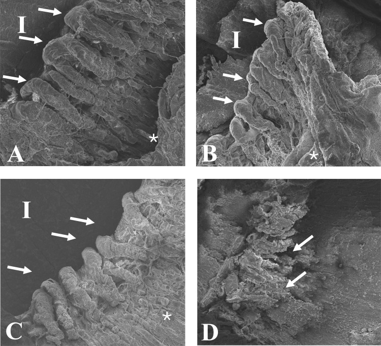
Results: SEM imaging revealed that both standard and Leos ECP resulted in blunting of the ciliary processes without injury to collateral tissues. In contrast, TCP-treated tissues showed significant structural disorganization extending to the iris and pars plana.
Conclusion: In this study, we demonstrate that both standard and novel Leos ECP techniques produce significantly less structural disruption to the ciliary processes compared to TCP. The Leos ECP system, with its enhanced imaging and automation features, may offer more clinical value through ease-of-use and improved consistency while still generating similar tissue effects to standard ECP. Additionally, these findings further validate the more targeted approach that ECP offers compared to TCP. However, no clinical outcomes were evaluated in this study, and further investigation is needed to determine how these findings translate to patient care.

 求助内容:
求助内容: 应助结果提醒方式:


