Qingxia Shen, Junqian Wang, Na Yao, Xiyan Niu, Mi Liu, Xiaohui Li
{"title":"Hepatoprotective effect of lotus leaf against non-alcoholic fatty liver disease in rats via alteration of AMPK/SIRT1 and Nrf2/HO-1 signaling pathway.","authors":"Qingxia Shen, Junqian Wang, Na Yao, Xiyan Niu, Mi Liu, Xiaohui Li","doi":"10.1590/acb407025","DOIUrl":null,"url":null,"abstract":"<p><strong>Purpose: </strong>In this study, we scrutinized the protective effect of lotus leaf (LF) against high-fat diet (HFD) induced liver injury in rats.</p><p><strong>Methods: </strong>The rats received the HFD for the induction of non-alcoholic fatty liver disease. Rats received the oral administration of LF (25, 50, and 100 mg/kg, b.w.). The insulin level, organ index, glucose level, hepatic, oxidative stress, lipid and cytokines parameters were measured. The different mRNA expression and histopathology were performed in the hepatic tissue.</p><p><strong>Results: </strong>LF treatment suppressed the insulin, glucose and HOMA-IR along with organ index (liver index and spleen index). LF treatment altered the level of liver parameters (aspartate aminotransferase, alanine aminotransferase, alkaline phosphatase, gamma-glutamyl transferase) and oxidative stress parameters in the serum, as well as the liver tissue. LF treatment altered the level of lipid parameters and fat parameters (total fat, perirenal fat, abdominal fat, epididymal fat); cytokines (tumor necrosis factor-α, interleukin-1β, interleukin-6, interleukin-10, interleukin-17, interleukin-33); HO-1, and Nrf2. LF treatment altered the mRNA expression of tumor necrosis factor-α, interleukin-1β, interleukin-6, interleukin-10, caspase-3, caspase-9, cytochrome C, cytochrome D, AMP-activated protein kinase (AMPK), sirtuin 1 (SIRT1), FRX-1, liver X Receptor alpha, fibronectin, matrix metalloproteinase-9, inducible nitric oxide synthase, and transforming growth factor-β 1 (TGF-β1). LF treatment suppressed the necrosis of hepatocytes with less inflammatory cell infiltration in the liver tissue along with alteration of liver injury score.</p><p><strong>Conclusion: </strong>The result showed the protective effect of LF against non-alcoholic fatty liver disease via activating the AMPK/SIRT1 and Nrf2/HO-1 pathway activation.</p>","PeriodicalId":93850,"journal":{"name":"Acta cirurgica brasileira","volume":"40 ","pages":"e407025"},"PeriodicalIF":1.3000,"publicationDate":"2025-08-25","publicationTypes":"Journal Article","fieldsOfStudy":null,"isOpenAccess":false,"openAccessPdf":"https://www.ncbi.nlm.nih.gov/pmc/articles/PMC12377701/pdf/","citationCount":"0","resultStr":null,"platform":"Semanticscholar","paperid":null,"PeriodicalName":"Acta cirurgica brasileira","FirstCategoryId":"1085","ListUrlMain":"https://doi.org/10.1590/acb407025","RegionNum":0,"RegionCategory":null,"ArticlePicture":[],"TitleCN":null,"AbstractTextCN":null,"PMCID":null,"EPubDate":"2025/1/1 0:00:00","PubModel":"eCollection","JCR":"","JCRName":"","Score":null,"Total":0}
引用次数: 0
Abstract
Purpose: In this study, we scrutinized the protective effect of lotus leaf (LF) against high-fat diet (HFD) induced liver injury in rats.
Methods: The rats received the HFD for the induction of non-alcoholic fatty liver disease. Rats received the oral administration of LF (25, 50, and 100 mg/kg, b.w.). The insulin level, organ index, glucose level, hepatic, oxidative stress, lipid and cytokines parameters were measured. The different mRNA expression and histopathology were performed in the hepatic tissue.
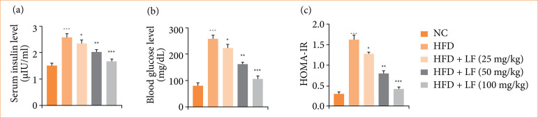
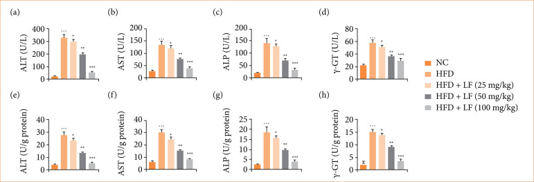
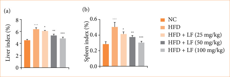
Results: LF treatment suppressed the insulin, glucose and HOMA-IR along with organ index (liver index and spleen index). LF treatment altered the level of liver parameters (aspartate aminotransferase, alanine aminotransferase, alkaline phosphatase, gamma-glutamyl transferase) and oxidative stress parameters in the serum, as well as the liver tissue. LF treatment altered the level of lipid parameters and fat parameters (total fat, perirenal fat, abdominal fat, epididymal fat); cytokines (tumor necrosis factor-α, interleukin-1β, interleukin-6, interleukin-10, interleukin-17, interleukin-33); HO-1, and Nrf2. LF treatment altered the mRNA expression of tumor necrosis factor-α, interleukin-1β, interleukin-6, interleukin-10, caspase-3, caspase-9, cytochrome C, cytochrome D, AMP-activated protein kinase (AMPK), sirtuin 1 (SIRT1), FRX-1, liver X Receptor alpha, fibronectin, matrix metalloproteinase-9, inducible nitric oxide synthase, and transforming growth factor-β 1 (TGF-β1). LF treatment suppressed the necrosis of hepatocytes with less inflammatory cell infiltration in the liver tissue along with alteration of liver injury score.
Conclusion: The result showed the protective effect of LF against non-alcoholic fatty liver disease via activating the AMPK/SIRT1 and Nrf2/HO-1 pathway activation.

 求助内容:
求助内容: 应助结果提醒方式:


