Imaging the mechanical properties of a developing embryonic tendon using Brillouin microscopy
IF 9.6
1区 医学
Q1 ENGINEERING, BIOMEDICAL
引用次数: 0
Abstract
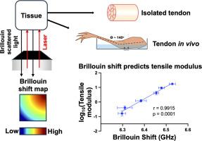
Tendon mechanical properties are critical for proper musculoskeletal movement. Yet, current understanding of how tendons develop their mechanical properties remains incomplete. Atomic force microscopy and tensile testing are used to characterize early-stage and late-stage embryonic tendon mechanical properties respectively, but both require contact with the tissue, which can alter or destroy tissue structure or integrity and render the tissue unsuitable for subsequent assays. Brillouin confocal microscopy is an emerging tool used to measure mechanical properties of tissues in an all-optical, contact-free, label-free manner. Here, Brillouin confocal microscopy was successfully implemented to measure chick embryo Achilles tendon mechanical properties, which were then validated by comparisons to previously published tensile modulus data. Significant changes in mechanical properties that occur with development and due to experimentally induced changes in extracellular matrix crosslinking were quantifiable. Brillouin microscopy also detected increasing mechanical heterogeneity of developing tendons. The potential to map the spatial distribution of the mechanical properties of developing extracellular matrix using Brillouin microscopy was demonstrated. Measurement of mechanical properties of the tendon within the limb, and sensitivity to changes in tendon strain imposed by changing ankle flexion demonstrated the potential for Brillouin microscopy to quantify tendon properties in vivo. Collectively, these results provide an exciting first study using Brillouin microscopy for quantitative and spatial mapping of embryonic tendon mechanical properties in an all-optical, label-free manner, which could lead to mechanistic discoveries not possible with conventional mechanical testing methodologies.
Statement of significance
Tendon injuries and disorders are problematic, as tendons lack the ability to regenerate, and current clinical solutions are insufficient. Knowledge gained from elucidating how embryonic tendons develop their mechanical properties could be used to inform the development of therapeutics to treat injured or abnormal tendons. However, conventional mechanical testing methods require challenging manipulations of small tissues and render tissues unusable for additional assays. Brillouin microscopy is a powerful technique that can image mechanical properties of tissues in a non-contact and label-free manner, which enables in vivo analysis of mechanical properties. In this study, we show that Brillouin microscopy can image mechanical properties of tendon in developing embryos, thus opening new avenues for investigation into the development of tendon mechanical properties.
用布里渊显微镜成像发育中的胚胎肌腱的力学特性。
肌腱的力学特性对肌肉骨骼的正常运动至关重要。然而,目前对肌腱如何发展其力学性能的理解仍然不完整。原子力显微镜和拉伸测试分别用于表征早期和晚期胚胎肌腱的力学特性,但两者都需要与组织接触,这可能会改变或破坏组织结构或完整性,并使组织不适合随后的分析。布里渊共聚焦显微镜是一种新兴的工具,用于测量组织的机械性能在全光学,无接触,无标签的方式。在这里,成功地使用布里渊共聚焦显微镜测量鸡胚胎跟腱的力学特性,然后通过与先前发表的拉伸模量数据进行比较来验证。随着发育和实验诱导的细胞外基质交联的变化,机械性能的显著变化是可量化的。布里渊显微镜还检测到发育中的肌腱的力学不均匀性增加。利用布里渊显微镜绘制发育中的细胞外基质的力学特性的空间分布的潜力得到了证明。肢体肌腱力学性能的测量,以及对踝关节屈曲变化引起的肌腱应变变化的敏感性,证明了布里渊显微镜在体内量化肌腱特性的潜力。总的来说,这些结果提供了一个令人兴奋的首次研究,使用布里渊显微镜以全光学、无标签的方式对胚胎肌腱的力学特性进行定量和空间测绘,这可能导致传统力学测试方法无法实现的力学发现。意义声明:肌腱损伤和紊乱是有问题的,因为肌腱缺乏再生能力,目前的临床解决方案不足。从阐明胚胎肌腱如何发展其机械特性中获得的知识可用于告知治疗受伤或异常肌腱的治疗方法的发展。然而,传统的机械测试方法需要对小组织进行具有挑战性的操作,并且使组织无法用于额外的分析。布里渊显微镜是一种强大的技术,它可以以非接触和无标签的方式对组织的机械特性进行成像,从而实现对机械特性的体内分析。在这项研究中,我们发现布里渊显微镜可以成像发育中的胚胎肌腱的力学特性,从而为研究肌腱力学特性的发展开辟了新的途径。
本文章由计算机程序翻译,如有差异,请以英文原文为准。
求助全文
约1分钟内获得全文
求助全文
来源期刊
Acta Biomaterialia
工程技术-材料科学:生物材料
CiteScore
16.80
自引率
3.10%
发文量
776
审稿时长
30 days
期刊介绍:
Acta Biomaterialia is a monthly peer-reviewed scientific journal published by Elsevier. The journal was established in January 2005. The editor-in-chief is W.R. Wagner (University of Pittsburgh). The journal covers research in biomaterials science, including the interrelationship of biomaterial structure and function from macroscale to nanoscale. Topical coverage includes biomedical and biocompatible materials.
 求助内容:
求助内容: 应助结果提醒方式:


