Mariola Krzyścin, Agnieszka Brodowska, Dominika Pietrzyk, Katarzyna Zając, Elżbieta Sowińska-Przepiera
{"title":"A Case Report of Tissue Mosaicism in 45,X0/46,XY: Diagnostic Complexity in a Newborn with Ambiguous Genitalia.","authors":"Mariola Krzyścin, Agnieszka Brodowska, Dominika Pietrzyk, Katarzyna Zając, Elżbieta Sowińska-Przepiera","doi":"10.3390/reports8030146","DOIUrl":null,"url":null,"abstract":"<p><p><b>Background and Clinical Significance</b>: The 45,X0/46,XY mosaic karyotype is categorized as a disorder of sex development and can lead to atypical sexual development. Latent mosaicism involving Y chromosomal segments may be much more prevalent than previously assumed, according to a growing number of findings. This primarily depends on how sensitive cytogenetic methods are-such as traditional karyotype screening, FISH methods, or molecular analyses. <b>Case Presentation</b>: We present the case of a 10-week-old infant with hermaphroditic external genitalia. During pregnancy, ultrasonography revealed severe fetal development difficulties, including severe widespread edema. An abnormal 45,X0/46,XY mosaic karyotype was discovered during a genetic amniocentesis conducted during the 16th week of pregnancy. The infant was born in average general condition at 39 + 6 weeks of gestation. Physical examination of the infant revealed features of facial dysmorphia, webbed neck, and hermaphroditic external genitalia. The testicle was palpable on the left side, but the gonad was absent on the right. Laboratory tests revealed a typical hormonal profile of the mini-puberty period in boys. Moreover, a hormone panel and thyroid ultrasound were performed; congenital hypothyroidism was diagnosed. Three separate independent sources of biological material were used in cytogenetic analysis to determine the karyotype: skin fibroblasts (to confirm tissue mosaicism), oral epithelial cells (FISH), and peripheral blood lymphocytes. It showed that a mosaic occurred very early in embryogenesis by confirming the existence of karyotypes 45,X and 46,XY in various tissues (mosaic tissue distribution). <b>Conclusions</b>: Tissue mosaicism should be compared to the analysis of tissues from other embryonic origins, including blood and oral tissue. Support for gender identity and treatment decisions, including the prediction of the future risk of gonadoblastoma, as well as multidisciplinary care, is necessary.</p>","PeriodicalId":74664,"journal":{"name":"Reports (MDPI)","volume":"8 3","pages":""},"PeriodicalIF":0.8000,"publicationDate":"2025-08-15","publicationTypes":"Journal Article","fieldsOfStudy":null,"isOpenAccess":false,"openAccessPdf":"https://www.ncbi.nlm.nih.gov/pmc/articles/PMC12371900/pdf/","citationCount":"0","resultStr":null,"platform":"Semanticscholar","paperid":null,"PeriodicalName":"Reports (MDPI)","FirstCategoryId":"1085","ListUrlMain":"https://doi.org/10.3390/reports8030146","RegionNum":0,"RegionCategory":null,"ArticlePicture":[],"TitleCN":null,"AbstractTextCN":null,"PMCID":null,"EPubDate":"","PubModel":"","JCR":"Q3","JCRName":"MEDICINE, GENERAL & INTERNAL","Score":null,"Total":0}
引用次数: 0
Abstract
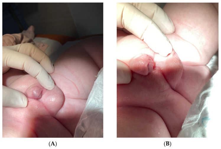
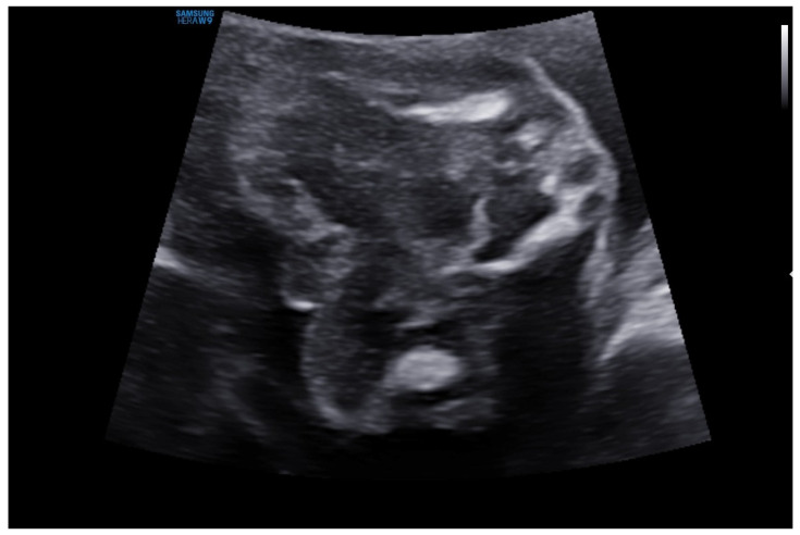
Background and Clinical Significance: The 45,X0/46,XY mosaic karyotype is categorized as a disorder of sex development and can lead to atypical sexual development. Latent mosaicism involving Y chromosomal segments may be much more prevalent than previously assumed, according to a growing number of findings. This primarily depends on how sensitive cytogenetic methods are-such as traditional karyotype screening, FISH methods, or molecular analyses. Case Presentation: We present the case of a 10-week-old infant with hermaphroditic external genitalia. During pregnancy, ultrasonography revealed severe fetal development difficulties, including severe widespread edema. An abnormal 45,X0/46,XY mosaic karyotype was discovered during a genetic amniocentesis conducted during the 16th week of pregnancy. The infant was born in average general condition at 39 + 6 weeks of gestation. Physical examination of the infant revealed features of facial dysmorphia, webbed neck, and hermaphroditic external genitalia. The testicle was palpable on the left side, but the gonad was absent on the right. Laboratory tests revealed a typical hormonal profile of the mini-puberty period in boys. Moreover, a hormone panel and thyroid ultrasound were performed; congenital hypothyroidism was diagnosed. Three separate independent sources of biological material were used in cytogenetic analysis to determine the karyotype: skin fibroblasts (to confirm tissue mosaicism), oral epithelial cells (FISH), and peripheral blood lymphocytes. It showed that a mosaic occurred very early in embryogenesis by confirming the existence of karyotypes 45,X and 46,XY in various tissues (mosaic tissue distribution). Conclusions: Tissue mosaicism should be compared to the analysis of tissues from other embryonic origins, including blood and oral tissue. Support for gender identity and treatment decisions, including the prediction of the future risk of gonadoblastoma, as well as multidisciplinary care, is necessary.

 求助内容:
求助内容: 应助结果提醒方式:


