Ana Maria Dumitrescu, Dragos Andrei Chiran, Cristinel Ionel Stan, Cringuta Mariana Paraschiv, Nicolaie Dobrin, Alexandru Chiriac, Maria Magdalena Leon, Lucia Corina Dima-Cozma, Cristina Gena Dascalu, Ana Marina Radulescu, Roxana Florentina Gavril, Anca Sava
{"title":"Comparing Antemortem CT-Angiography Data with Autopsy Findings in Regard to Anterior Communicating Artery Aneurysms.","authors":"Ana Maria Dumitrescu, Dragos Andrei Chiran, Cristinel Ionel Stan, Cringuta Mariana Paraschiv, Nicolaie Dobrin, Alexandru Chiriac, Maria Magdalena Leon, Lucia Corina Dima-Cozma, Cristina Gena Dascalu, Ana Marina Radulescu, Roxana Florentina Gavril, Anca Sava","doi":"10.3390/neurosci6030081","DOIUrl":null,"url":null,"abstract":"<p><strong>Background: </strong>The literature shows that anterior communicating artery (AcoA) aneurysms are the most common intracranial aneurysms. To date, there has only been one postmortem study focused on the correlations between autopsy findings and imaging results in cases of intracranial aneurysms associated with anatomical variants of the circle of Willis (CW).</p><p><strong>Methods: </strong>We investigated the anatomical variants of the CW associated with the occurrence and rupture of AcoA aneurysms by performing comparative analyses, in the same patients, of postmortem autopsy data with antemortem computed tomography-angiography (CTA) results obtained in the first 48 h after the onset of subarachnoid hemorrhage. Our retrospective observational study identified the anatomical variants of the CW at autopsy in 16 deceased adult Romanian patients with AcoA aneurysms over a 12-year period (2010-2022).</p><p><strong>Results: </strong>The autopsy findings revealed that the AcoA ruptured aneurysms had a mean external diameter of 9.50 mm, and 71.4% of them presented three or four anatomical variants inside the same CW. The initial antemortem CTA examination correctly located the AcoA aneurysms in all cases (100%), and an anatomical variant of the CW was only noted in 18.75% of patients. The final postmortem re-analyzed the same CTA images identified in all cases (100%), focusing on both the AcoA aneurysm and all anatomical variants of the CW found during the autopsies.</p><p><strong>Conclusions: </strong>Although it was previously thought that the occurrence of AcoA aneurysms is related only to the hemodynamic changes induced by the nearby arterial anatomical variants, we identified the simultaneous involvement of at least one hypoplastic artery and one or two PCA fetal-type anatomical variants that were located in both the anterior and posterior parts of the CW. Furthermore, if sufficient time is devoted to the CT-angiography analysis and interpretation of the images, anatomical variants of the circle of Willis associated with AcoA aneurysms can be identified as accurately as they are in invasive postmortem autopsy examinations.</p>","PeriodicalId":74294,"journal":{"name":"NeuroSci","volume":"6 3","pages":""},"PeriodicalIF":2.0000,"publicationDate":"2025-08-18","publicationTypes":"Journal Article","fieldsOfStudy":null,"isOpenAccess":false,"openAccessPdf":"https://www.ncbi.nlm.nih.gov/pmc/articles/PMC12371958/pdf/","citationCount":"0","resultStr":null,"platform":"Semanticscholar","paperid":null,"PeriodicalName":"NeuroSci","FirstCategoryId":"1085","ListUrlMain":"https://doi.org/10.3390/neurosci6030081","RegionNum":0,"RegionCategory":null,"ArticlePicture":[],"TitleCN":null,"AbstractTextCN":null,"PMCID":null,"EPubDate":"","PubModel":"","JCR":"Q3","JCRName":"CLINICAL NEUROLOGY","Score":null,"Total":0}
引用次数: 0
Abstract
Background: The literature shows that anterior communicating artery (AcoA) aneurysms are the most common intracranial aneurysms. To date, there has only been one postmortem study focused on the correlations between autopsy findings and imaging results in cases of intracranial aneurysms associated with anatomical variants of the circle of Willis (CW).
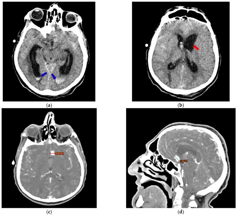
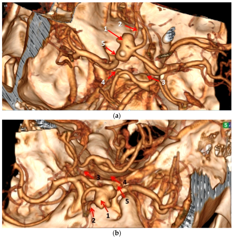
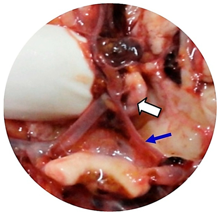
Methods: We investigated the anatomical variants of the CW associated with the occurrence and rupture of AcoA aneurysms by performing comparative analyses, in the same patients, of postmortem autopsy data with antemortem computed tomography-angiography (CTA) results obtained in the first 48 h after the onset of subarachnoid hemorrhage. Our retrospective observational study identified the anatomical variants of the CW at autopsy in 16 deceased adult Romanian patients with AcoA aneurysms over a 12-year period (2010-2022).
Results: The autopsy findings revealed that the AcoA ruptured aneurysms had a mean external diameter of 9.50 mm, and 71.4% of them presented three or four anatomical variants inside the same CW. The initial antemortem CTA examination correctly located the AcoA aneurysms in all cases (100%), and an anatomical variant of the CW was only noted in 18.75% of patients. The final postmortem re-analyzed the same CTA images identified in all cases (100%), focusing on both the AcoA aneurysm and all anatomical variants of the CW found during the autopsies.
Conclusions: Although it was previously thought that the occurrence of AcoA aneurysms is related only to the hemodynamic changes induced by the nearby arterial anatomical variants, we identified the simultaneous involvement of at least one hypoplastic artery and one or two PCA fetal-type anatomical variants that were located in both the anterior and posterior parts of the CW. Furthermore, if sufficient time is devoted to the CT-angiography analysis and interpretation of the images, anatomical variants of the circle of Willis associated with AcoA aneurysms can be identified as accurately as they are in invasive postmortem autopsy examinations.

 求助内容:
求助内容: 应助结果提醒方式:


