Nurfarhanah Bte Syed Sulaiman, Sofiah M Y Sng, Khurshid Z Merchant, Lee Ping Ng, David C Y Low, Wan Tew Seow, Sharon Y Y Low
{"title":"Identifying Angiogenic Factors in Pediatric Choroid Plexus Papillomas.","authors":"Nurfarhanah Bte Syed Sulaiman, Sofiah M Y Sng, Khurshid Z Merchant, Lee Ping Ng, David C Y Low, Wan Tew Seow, Sharon Y Y Low","doi":"10.3390/neurosci6030076","DOIUrl":null,"url":null,"abstract":"<p><p>(1) Background: Choroid plexus papillomas (CPPs) are rare brain tumors that tend to occur in very young children. Mechanisms of CPP development remain unelucidated. Separately, the process of angiogenesis has been implicated in other primary brain tumors. We hypothesize that angiogenesis is a hallmark of CPP biology. This study aims to identify and validate angiogenic factors in CPPs. (2) Methods: Cerebrospinal fluid (CSF) and CPP tumor samples are collected. A multiplex immunoassay panel is used to identify differentially expressed cytokines in the CSF samples. Concurrently, patient-derived primary cell cultures and their supernatants are derived from CPP samples. Targeted proteome blot arrays and human umbilical vein endothelial cell (HUVEC) angiogenesis assays are used for validation studies. (3) Results: CSF profiling showed higher expressions of VEGF-A, MCP-1, MMP-1, TNF-α, and CD40L in CPP patient samples versus non-tumor controls. Next, assessment via online protein-protein network platforms reports that these cytokines are associated with endothelial cell regulation. Using an angiogenesis-focused approach, CPP-derived cell lines and supernatants showed similarly higher expressions of VEGF, MCP-1, and MMP-1. Next, sprouting of nodes and tubule formation were observed in HUVEC angiogenesis assay cultures when conditioned CPP cell culture media was added. (4) Conclusions: This proof-of-concept study demonstrates potential to explore angiogenesis in CPP.</p>","PeriodicalId":74294,"journal":{"name":"NeuroSci","volume":"6 3","pages":""},"PeriodicalIF":2.0000,"publicationDate":"2025-08-11","publicationTypes":"Journal Article","fieldsOfStudy":null,"isOpenAccess":false,"openAccessPdf":"https://www.ncbi.nlm.nih.gov/pmc/articles/PMC12372068/pdf/","citationCount":"0","resultStr":null,"platform":"Semanticscholar","paperid":null,"PeriodicalName":"NeuroSci","FirstCategoryId":"1085","ListUrlMain":"https://doi.org/10.3390/neurosci6030076","RegionNum":0,"RegionCategory":null,"ArticlePicture":[],"TitleCN":null,"AbstractTextCN":null,"PMCID":null,"EPubDate":"","PubModel":"","JCR":"Q3","JCRName":"CLINICAL NEUROLOGY","Score":null,"Total":0}
引用次数: 0
Abstract
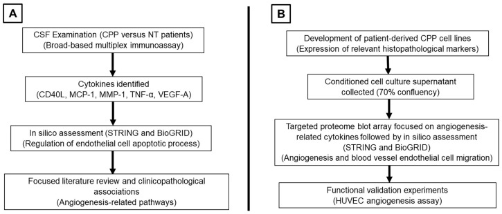
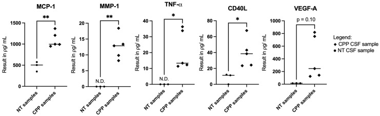
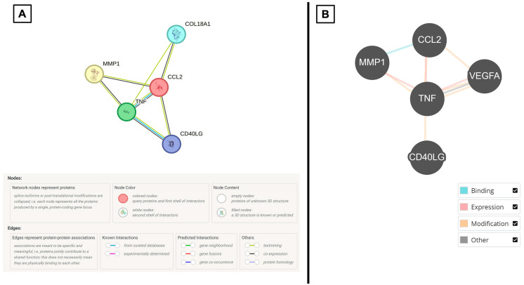
(1) Background: Choroid plexus papillomas (CPPs) are rare brain tumors that tend to occur in very young children. Mechanisms of CPP development remain unelucidated. Separately, the process of angiogenesis has been implicated in other primary brain tumors. We hypothesize that angiogenesis is a hallmark of CPP biology. This study aims to identify and validate angiogenic factors in CPPs. (2) Methods: Cerebrospinal fluid (CSF) and CPP tumor samples are collected. A multiplex immunoassay panel is used to identify differentially expressed cytokines in the CSF samples. Concurrently, patient-derived primary cell cultures and their supernatants are derived from CPP samples. Targeted proteome blot arrays and human umbilical vein endothelial cell (HUVEC) angiogenesis assays are used for validation studies. (3) Results: CSF profiling showed higher expressions of VEGF-A, MCP-1, MMP-1, TNF-α, and CD40L in CPP patient samples versus non-tumor controls. Next, assessment via online protein-protein network platforms reports that these cytokines are associated with endothelial cell regulation. Using an angiogenesis-focused approach, CPP-derived cell lines and supernatants showed similarly higher expressions of VEGF, MCP-1, and MMP-1. Next, sprouting of nodes and tubule formation were observed in HUVEC angiogenesis assay cultures when conditioned CPP cell culture media was added. (4) Conclusions: This proof-of-concept study demonstrates potential to explore angiogenesis in CPP.

 求助内容:
求助内容: 应助结果提醒方式:


