N V Tarbaeva, A V Manaev, K V Ivashchenko, N M Platonova, D G Beltsevich, N V Pachuashvili, L S Urusova, N G Mokrysheva
{"title":"The value of CT texture analysis in predicting mitotic activity and morphological variants of adrenocortical carcinoma.","authors":"N V Tarbaeva, A V Manaev, K V Ivashchenko, N M Platonova, D G Beltsevich, N V Pachuashvili, L S Urusova, N G Mokrysheva","doi":"10.3389/fradi.2025.1635425","DOIUrl":null,"url":null,"abstract":"<p><strong>Introduction: </strong>Adrenocortical carcinoma presents significant diagnostic challenges due to its histological heterogeneity and variable clinical behavior. This study aimed to evaluate the diagnostic value of radiomic features in predicting mitotic activity (low/high-grade) and morphological variants (conventional, oncocytic, myxoid) of adrenocortical carcinoma.</p><p><strong>Materials and methods: </strong>A retrospective analysis of 32 patients with histologically confirmed ACC (18 conventional, 9 oncocytic and 5 myxoid cases) was performed, with mitotic data available for 25 cases (13 low-grade and 12 high-grade cases). Radiomic features including Gray-Level Co-occurrence Matrix (GLCM), Run-Length (GLRLM), Size-Zone (GLSZM), Dependence (GLDM), Neighboring-Tone (NGTDM) and first order features were extracted from four-phase CT using PyRadiomics after manual 3D segmentation. Statistical analysis included Mann-Whitney <i>U</i>, Kruskal-Wallis tests, ROC curve (AUC, sensitivity, specificity) and PPV, NPV assessment.</p><p><strong>Results: </strong>Our analysis demonstrated statistically significant differences between tumor grades with firstorder_Skewness (AUC = 0.924, 95% CI: 0.819-0.986; <i>p</i> = 0.005) showing high predictive performance in the venous phase. Radiomic features did not show statistically significant differences between morphological variants of ACC after adjustment for multiple comparisons.</p><p><strong>Conclusion: </strong>Our results confirm the value of CT radiomics for preoperative stratification of ACC grade, but the question of differentiation of morphological variants remains unresolved and requires further validation in larger cohorts.</p>","PeriodicalId":73101,"journal":{"name":"Frontiers in radiology","volume":"5 ","pages":"1635425"},"PeriodicalIF":2.3000,"publicationDate":"2025-08-07","publicationTypes":"Journal Article","fieldsOfStudy":null,"isOpenAccess":false,"openAccessPdf":"https://www.ncbi.nlm.nih.gov/pmc/articles/PMC12367671/pdf/","citationCount":"0","resultStr":null,"platform":"Semanticscholar","paperid":null,"PeriodicalName":"Frontiers in radiology","FirstCategoryId":"1085","ListUrlMain":"https://doi.org/10.3389/fradi.2025.1635425","RegionNum":0,"RegionCategory":null,"ArticlePicture":[],"TitleCN":null,"AbstractTextCN":null,"PMCID":null,"EPubDate":"2025/1/1 0:00:00","PubModel":"eCollection","JCR":"","JCRName":"","Score":null,"Total":0}
引用次数: 0
Abstract
Introduction: Adrenocortical carcinoma presents significant diagnostic challenges due to its histological heterogeneity and variable clinical behavior. This study aimed to evaluate the diagnostic value of radiomic features in predicting mitotic activity (low/high-grade) and morphological variants (conventional, oncocytic, myxoid) of adrenocortical carcinoma.
Materials and methods: A retrospective analysis of 32 patients with histologically confirmed ACC (18 conventional, 9 oncocytic and 5 myxoid cases) was performed, with mitotic data available for 25 cases (13 low-grade and 12 high-grade cases). Radiomic features including Gray-Level Co-occurrence Matrix (GLCM), Run-Length (GLRLM), Size-Zone (GLSZM), Dependence (GLDM), Neighboring-Tone (NGTDM) and first order features were extracted from four-phase CT using PyRadiomics after manual 3D segmentation. Statistical analysis included Mann-Whitney U, Kruskal-Wallis tests, ROC curve (AUC, sensitivity, specificity) and PPV, NPV assessment.
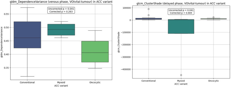
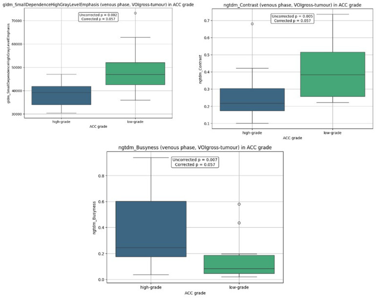
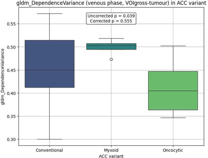
Results: Our analysis demonstrated statistically significant differences between tumor grades with firstorder_Skewness (AUC = 0.924, 95% CI: 0.819-0.986; p = 0.005) showing high predictive performance in the venous phase. Radiomic features did not show statistically significant differences between morphological variants of ACC after adjustment for multiple comparisons.
Conclusion: Our results confirm the value of CT radiomics for preoperative stratification of ACC grade, but the question of differentiation of morphological variants remains unresolved and requires further validation in larger cohorts.

 求助内容:
求助内容: 应助结果提醒方式:


