Tamer R Hage, Edward J Kelly, Eriks Ziedins, Babita Parajuli, Cameron S D'Orio, David M Burmeister, Lauren Moffatt, Jeffrey W Shupp, Bonnie C Carney
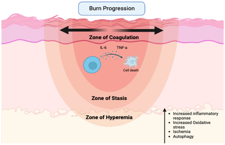
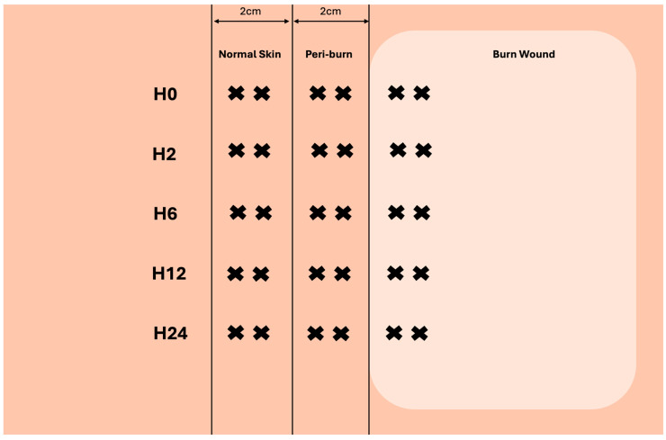
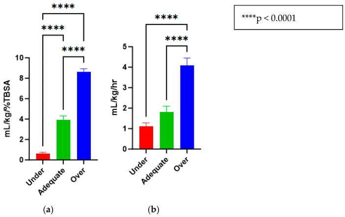
{"title":"Pilot Study on Resuscitation Volume's Effect on Perfusion and Inflammatory Cytokine Expression in Peri-Burn Skin: Implications for Burn Conversion.","authors":"Tamer R Hage, Edward J Kelly, Eriks Ziedins, Babita Parajuli, Cameron S D'Orio, David M Burmeister, Lauren Moffatt, Jeffrey W Shupp, Bonnie C Carney","doi":"10.3390/ebj6030042","DOIUrl":null,"url":null,"abstract":"<p><p>Fluid resuscitation after thermal injury is paramount to avoid burn shock and restore organ perfusion. Both over- and under-resuscitation can lead to unintended consequences affecting patient outcomes. While many studies have examined systemic effects, limited data exist on how fluid resuscitation impacts burn wound progression in the acute period. Furthermore, the mechanisms underlying burn wound progression remain not fully understood. This study used a swine model to investigate how varying resuscitation levels affect peri-burn wound dynamics. Twenty-seven female Yorkshire pigs were anesthetized, subjected to 40% total body surface area burn and 15% hemorrhage, then randomized (<i>n</i> = 9) to receive decision-support-driven (adequate, 2-4 mL/kg/%TBSA), fluid-withholding (under, <1 mL/kg/%TBSA), or high-constant-rate (over, >>4 mL/kg/%TBSA) resuscitation. Pigs were monitored for 24 h in an intensive care setting prior to necropsy. Laser Doppler Imaging (LDI) was conducted pre-burn and at 2, 6, 12, and 24 h post burn to assess perfusion. Biopsies were taken from burn, peri-burn (within 2 cm), and normal skin. RNA was isolated at 24 h for the qRT-PCR analysis of IL-6, CXCL8, and IFN-γ. At hour 2, LDI revealed increased peri-burn perfusion in over-resuscitated animals vs. under-resuscitated animals (<i>p</i> = 0.0499). At hour 24, IL-6 (<i>p</i> = 0.0220) and IFN-γ (<i>p</i> = 0.0253) were elevated in over-resuscitated peri-burn skin. CXCL8 showed no significant change. TUNEL staining revealed increased apoptosis in over- and under-resuscitated peri-burn skin. Differences in perfusion and cytokine expression based on resuscitation strategy suggest that fluid levels may influence burn wound progression.</p>","PeriodicalId":72961,"journal":{"name":"European burn journal","volume":"6 3","pages":""},"PeriodicalIF":1.2000,"publicationDate":"2025-07-28","publicationTypes":"Journal Article","fieldsOfStudy":null,"isOpenAccess":false,"openAccessPdf":"https://www.ncbi.nlm.nih.gov/pmc/articles/PMC12372052/pdf/","citationCount":"0","resultStr":null,"platform":"Semanticscholar","paperid":null,"PeriodicalName":"European burn journal","FirstCategoryId":"1085","ListUrlMain":"https://doi.org/10.3390/ebj6030042","RegionNum":0,"RegionCategory":null,"ArticlePicture":[],"TitleCN":null,"AbstractTextCN":null,"PMCID":null,"EPubDate":"","PubModel":"","JCR":"Q4","JCRName":"CRITICAL CARE MEDICINE","Score":null,"Total":0}
引用次数: 0
Abstract
Fluid resuscitation after thermal injury is paramount to avoid burn shock and restore organ perfusion. Both over- and under-resuscitation can lead to unintended consequences affecting patient outcomes. While many studies have examined systemic effects, limited data exist on how fluid resuscitation impacts burn wound progression in the acute period. Furthermore, the mechanisms underlying burn wound progression remain not fully understood. This study used a swine model to investigate how varying resuscitation levels affect peri-burn wound dynamics. Twenty-seven female Yorkshire pigs were anesthetized, subjected to 40% total body surface area burn and 15% hemorrhage, then randomized (n = 9) to receive decision-support-driven (adequate, 2-4 mL/kg/%TBSA), fluid-withholding (under, <1 mL/kg/%TBSA), or high-constant-rate (over, >>4 mL/kg/%TBSA) resuscitation. Pigs were monitored for 24 h in an intensive care setting prior to necropsy. Laser Doppler Imaging (LDI) was conducted pre-burn and at 2, 6, 12, and 24 h post burn to assess perfusion. Biopsies were taken from burn, peri-burn (within 2 cm), and normal skin. RNA was isolated at 24 h for the qRT-PCR analysis of IL-6, CXCL8, and IFN-γ. At hour 2, LDI revealed increased peri-burn perfusion in over-resuscitated animals vs. under-resuscitated animals (p = 0.0499). At hour 24, IL-6 (p = 0.0220) and IFN-γ (p = 0.0253) were elevated in over-resuscitated peri-burn skin. CXCL8 showed no significant change. TUNEL staining revealed increased apoptosis in over- and under-resuscitated peri-burn skin. Differences in perfusion and cytokine expression based on resuscitation strategy suggest that fluid levels may influence burn wound progression.

 求助内容:
求助内容: 应助结果提醒方式:


