Yssel Mendoza-Mari, Marija Stojanovic, Dan E Miulli, Devendra K Agrawal
{"title":"Microglial Response to Inflammatory Stimuli Under Electromagnetic Field Exposure.","authors":"Yssel Mendoza-Mari, Marija Stojanovic, Dan E Miulli, Devendra K Agrawal","doi":"10.26502/acbr.50170467","DOIUrl":null,"url":null,"abstract":"<p><p>Microglial cells constitute the largest number of non-neuronal cells in the brain. As part of their immune surveillance function, they are responsible for detecting the presence of both external and internal danger signals, stimulating a defense response through the release of pro-inflammatory cytokines. Once the damage is controlled, microglia stimulate a reparative response that allows tissue homeostasis to be maintained. When this balance is not physiologically achieved, the use of drugs or other non-pharmacological therapies is needed to promote tissue repair and prevent the appearance of complications secondary to the primary damage. In the particular case of traumatic brain injury (TBI), the application of low frequency electromagnetic field (EMF) has proven very helpful in reducing the levels of inflammatory mediators. In the present study we investigated the effect of EMF in an \"<i>in vitro</i>\" model of tumor necrosis factor alpha (TNF-α)-induced neuroinflammation. Human microglial cells (HMC3) were treated with TNF-α (50 ng/mL) and, after 20 minutes, were exposed to 2.5 or 5 Hz EMF for 3 min. The effect of both treatments on survival, migration capacity and transcriptional expression of M1/M2 phenotypic markers was evaluated at 6, 24 and 48 hours. The exposure to EMF increased the survival rate of cells incubated with high doses of TNF-α and significantly reduced the migration rate of TNF-α treated cells. The analysis of expression patterns in different time points showed that EMF promoted the expression of M1 and M2 phenotypic markers in a time-dependent manner, suggesting a stimulating effect on the phagocytic capacity of microglial cells. Further studies are necessary to fully characterize the effects of EMF on the function of primary microglial cells within a brain injury-like microenvironment.</p>","PeriodicalId":72279,"journal":{"name":"Archives of clinical and biomedical research","volume":"9 4","pages":"304-315"},"PeriodicalIF":0.0000,"publicationDate":"2025-01-01","publicationTypes":"Journal Article","fieldsOfStudy":null,"isOpenAccess":false,"openAccessPdf":"https://www.ncbi.nlm.nih.gov/pmc/articles/PMC12372985/pdf/","citationCount":"0","resultStr":null,"platform":"Semanticscholar","paperid":null,"PeriodicalName":"Archives of clinical and biomedical research","FirstCategoryId":"1085","ListUrlMain":"https://doi.org/10.26502/acbr.50170467","RegionNum":0,"RegionCategory":null,"ArticlePicture":[],"TitleCN":null,"AbstractTextCN":null,"PMCID":null,"EPubDate":"2025/6/30 0:00:00","PubModel":"Epub","JCR":"","JCRName":"","Score":null,"Total":0}
引用次数: 0
Abstract
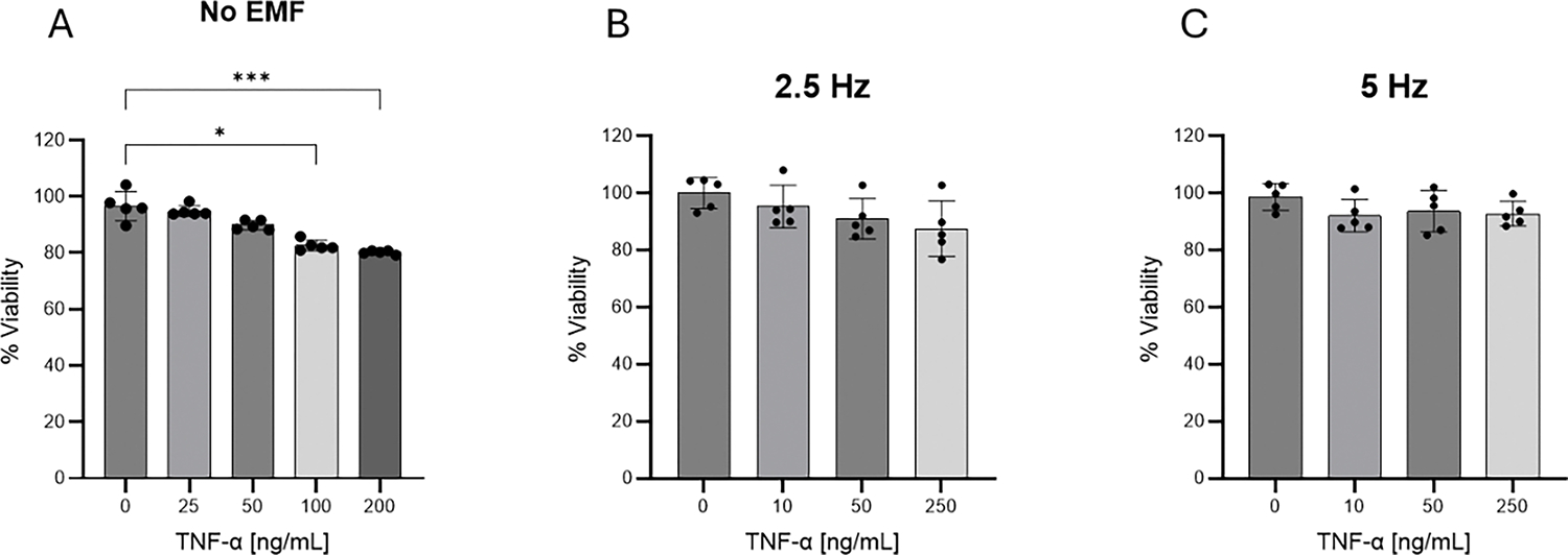
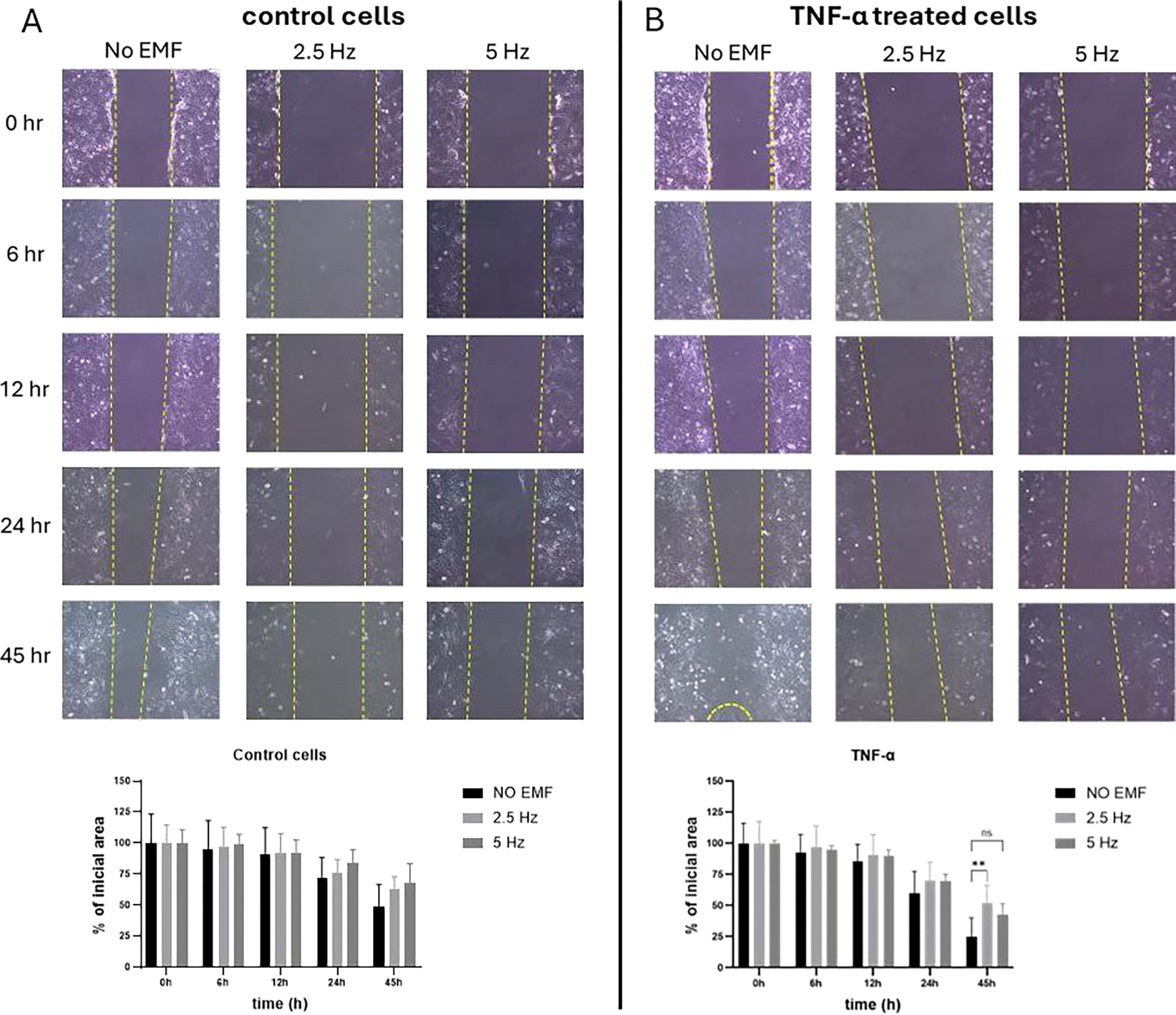
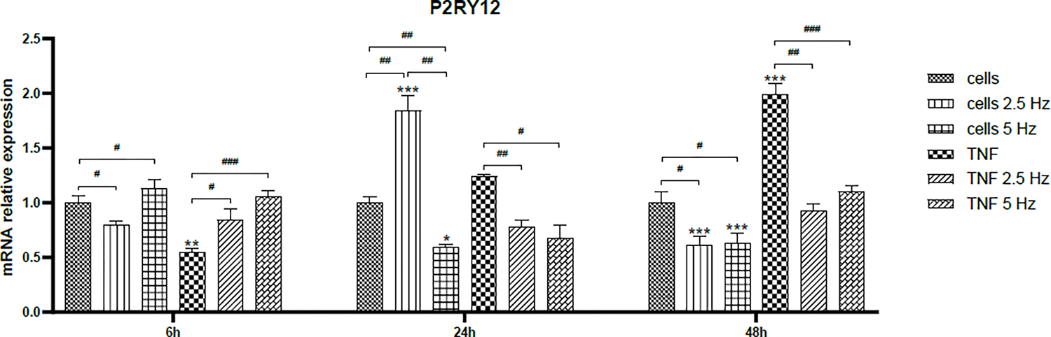
Microglial cells constitute the largest number of non-neuronal cells in the brain. As part of their immune surveillance function, they are responsible for detecting the presence of both external and internal danger signals, stimulating a defense response through the release of pro-inflammatory cytokines. Once the damage is controlled, microglia stimulate a reparative response that allows tissue homeostasis to be maintained. When this balance is not physiologically achieved, the use of drugs or other non-pharmacological therapies is needed to promote tissue repair and prevent the appearance of complications secondary to the primary damage. In the particular case of traumatic brain injury (TBI), the application of low frequency electromagnetic field (EMF) has proven very helpful in reducing the levels of inflammatory mediators. In the present study we investigated the effect of EMF in an "in vitro" model of tumor necrosis factor alpha (TNF-α)-induced neuroinflammation. Human microglial cells (HMC3) were treated with TNF-α (50 ng/mL) and, after 20 minutes, were exposed to 2.5 or 5 Hz EMF for 3 min. The effect of both treatments on survival, migration capacity and transcriptional expression of M1/M2 phenotypic markers was evaluated at 6, 24 and 48 hours. The exposure to EMF increased the survival rate of cells incubated with high doses of TNF-α and significantly reduced the migration rate of TNF-α treated cells. The analysis of expression patterns in different time points showed that EMF promoted the expression of M1 and M2 phenotypic markers in a time-dependent manner, suggesting a stimulating effect on the phagocytic capacity of microglial cells. Further studies are necessary to fully characterize the effects of EMF on the function of primary microglial cells within a brain injury-like microenvironment.

 求助内容:
求助内容: 应助结果提醒方式:


