{"title":"Tracheal mast cell tumor in a dog - surgical approach and diagnosis.","authors":"Fernanda Rezende Souza, Karen Yumi Ribeiro Nakagaki, Fernanda Freitas Miranda, Leonardo Dias Mamão, Geovanni Dantas Cassali","doi":"10.29374/2527-2179.bjvm000525","DOIUrl":null,"url":null,"abstract":"<p><p>Mast cell tumors (MCTs) are a type of cutaneous neoplasm prevalent in canines. Although less frequent, such neoplasms can involve other anatomical sites with no skin involvement, such as the trachea. The objective of the present case report is to describe the clinical, surgical, histopathological, and immunohistochemical features of a tracheal MCT in a dog. An 8-y-old, mixed-breed, male dog showed signs of dyspnea, coughing and choking. Tracheobronchoscopy revealed a mass in the cervical part of the trachea, almost completely obstructing its lumen. Surgery was performed for removal of the mass and part of the tracheal rings. Histologically, the trachea showed transmural thickening with a round cell neoplastic proliferation. Extracutaneous mast cell tumor was confirmed by toluidine blue staining. Immunohistochemistry was performed for c-KIT with KIT-staining II and Ki67 >23 cells/grid (and 73.2% positive cells). The dog exhibited no postoperative complications. A local recurrence occurred four months after surgery and the animal's general condition deteriorated, which led to the patient's euthanasia. Although rare, mast cell tumors should be considered in the differential diagnosis of dogs with extracutaneous nodules and masses.</p>","PeriodicalId":72458,"journal":{"name":"Brazilian journal of veterinary medicine","volume":"47 ","pages":"e000525"},"PeriodicalIF":0.0000,"publicationDate":"2025-08-19","publicationTypes":"Journal Article","fieldsOfStudy":null,"isOpenAccess":false,"openAccessPdf":"https://www.ncbi.nlm.nih.gov/pmc/articles/PMC12379833/pdf/","citationCount":"0","resultStr":null,"platform":"Semanticscholar","paperid":null,"PeriodicalName":"Brazilian journal of veterinary medicine","FirstCategoryId":"1085","ListUrlMain":"https://doi.org/10.29374/2527-2179.bjvm000525","RegionNum":0,"RegionCategory":null,"ArticlePicture":[],"TitleCN":null,"AbstractTextCN":null,"PMCID":null,"EPubDate":"2025/1/1 0:00:00","PubModel":"eCollection","JCR":"","JCRName":"","Score":null,"Total":0}
引用次数: 0
Abstract
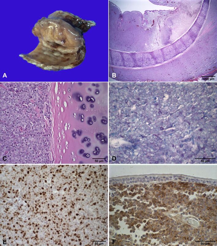
Mast cell tumors (MCTs) are a type of cutaneous neoplasm prevalent in canines. Although less frequent, such neoplasms can involve other anatomical sites with no skin involvement, such as the trachea. The objective of the present case report is to describe the clinical, surgical, histopathological, and immunohistochemical features of a tracheal MCT in a dog. An 8-y-old, mixed-breed, male dog showed signs of dyspnea, coughing and choking. Tracheobronchoscopy revealed a mass in the cervical part of the trachea, almost completely obstructing its lumen. Surgery was performed for removal of the mass and part of the tracheal rings. Histologically, the trachea showed transmural thickening with a round cell neoplastic proliferation. Extracutaneous mast cell tumor was confirmed by toluidine blue staining. Immunohistochemistry was performed for c-KIT with KIT-staining II and Ki67 >23 cells/grid (and 73.2% positive cells). The dog exhibited no postoperative complications. A local recurrence occurred four months after surgery and the animal's general condition deteriorated, which led to the patient's euthanasia. Although rare, mast cell tumors should be considered in the differential diagnosis of dogs with extracutaneous nodules and masses.


 求助内容:
求助内容: 应助结果提醒方式:


