{"title":"Pre-lacrimal window maxillary sinus surgery: a computed tomography analysis and classification.","authors":"Mohammad Waheed El-Anwar, Mohamed Kamel Alawady, Mohamed Alshawadfy, Mohamed Mohamed Rabea, Atef Hussein Elsayed","doi":"10.7181/acfs.2025.0023","DOIUrl":null,"url":null,"abstract":"<p><strong>Background: </strong>The pre-lacrimal window (PLW) approach is a promising technique for accessing otherwise inaccessible maxillary sinus lesions. The objective of this study was to determine the computed tomography (CT) dimensions, measurements, and grading of the PLW.</p><p><strong>Methods: </strong>One hundred paranasal CT scans were included in the study. For all subjects, axial images were obtained, and multiplanar reformats were used to obtain detailed views in the coronal and sagittal planes. The width of the PLW, the width of the nasolacrimal duct (NLD), and the angle between the axis of the NLD and the hard palate were measured and graded.</p><p><strong>Results: </strong>In 100 CT scans (200 sides), the mean PLW width was 5.6± 2.4 mm (range, 0-11.15 mm), the mean NLD width was 6.38± 1.84 mm (range, 1-11 mm), and the mean angle between the axis of the NLD and the hard palate was 68.6°± 6.77° (range, 54°-83°). There were no significant differences between sides or genders for any of the measurements.</p><p><strong>Conclusion: </strong>The CT dimensions of the PLW should be carefully evaluated when considering different endoscopic approaches to, or through, the anterior aspect of the maxillary sinus. The current study enhances surgeon and radiologist awareness of PLW measurements and their variations, ultimately improving the application of the PLW approach.</p>","PeriodicalId":52238,"journal":{"name":"Archives of Craniofacial Surgery","volume":"26 4","pages":"141-146"},"PeriodicalIF":0.0000,"publicationDate":"2025-08-01","publicationTypes":"Journal Article","fieldsOfStudy":null,"isOpenAccess":false,"openAccessPdf":"https://www.ncbi.nlm.nih.gov/pmc/articles/PMC12415367/pdf/","citationCount":"0","resultStr":null,"platform":"Semanticscholar","paperid":null,"PeriodicalName":"Archives of Craniofacial Surgery","FirstCategoryId":"1085","ListUrlMain":"https://doi.org/10.7181/acfs.2025.0023","RegionNum":0,"RegionCategory":null,"ArticlePicture":[],"TitleCN":null,"AbstractTextCN":null,"PMCID":null,"EPubDate":"2025/8/20 0:00:00","PubModel":"Epub","JCR":"Q2","JCRName":"Medicine","Score":null,"Total":0}
引用次数: 0
Abstract
Background: The pre-lacrimal window (PLW) approach is a promising technique for accessing otherwise inaccessible maxillary sinus lesions. The objective of this study was to determine the computed tomography (CT) dimensions, measurements, and grading of the PLW.
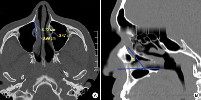
Methods: One hundred paranasal CT scans were included in the study. For all subjects, axial images were obtained, and multiplanar reformats were used to obtain detailed views in the coronal and sagittal planes. The width of the PLW, the width of the nasolacrimal duct (NLD), and the angle between the axis of the NLD and the hard palate were measured and graded.
Results: In 100 CT scans (200 sides), the mean PLW width was 5.6± 2.4 mm (range, 0-11.15 mm), the mean NLD width was 6.38± 1.84 mm (range, 1-11 mm), and the mean angle between the axis of the NLD and the hard palate was 68.6°± 6.77° (range, 54°-83°). There were no significant differences between sides or genders for any of the measurements.
Conclusion: The CT dimensions of the PLW should be carefully evaluated when considering different endoscopic approaches to, or through, the anterior aspect of the maxillary sinus. The current study enhances surgeon and radiologist awareness of PLW measurements and their variations, ultimately improving the application of the PLW approach.

 求助内容:
求助内容: 应助结果提醒方式:


