Zi Qiang Glen Liau, Wai Keong Ryan Loke, Danakkrisna Vachalam S/O Rangasamie, Yu Liu
{"title":"Novel all-intra-incisional pin placement technique in robotic total knee arthroplasty: a safer alternative.","authors":"Zi Qiang Glen Liau, Wai Keong Ryan Loke, Danakkrisna Vachalam S/O Rangasamie, Yu Liu","doi":"10.1186/s42836-025-00329-8","DOIUrl":null,"url":null,"abstract":"<p><strong>Introduction: </strong>Major robotic systems for total knee replacements necessitate the use of array pins in the tibia and femur. These extra-incisional pins are placed away from the primary incision and may be associated with soft tissue complications and peri-prosthetic fractures. There is currently no standardized, reproducible method for reliably placing pins in the femur and tibia metaphyses. We have developed an all-intra-incisional pin method within the primary incision. This paper aims to describe our technique, analyse the proximity of the pins to the implants, and study complications arising from both techniques.</p><p><strong>Methods: </strong>A total of 102 robotic-assisted total knee arthroplasties were performed using the ROSA, MAKO, and CORI systems. Patient charts were reviewed for their age, gender, body mass index, and ethnicity. Post-operative day zero radiographs of the operated knee were used for measurements in anteroposterior and lateral views, with X-ray magnifications taken into consideration.</p><p><strong>Results: </strong>Our study demonstrates that intra-incisional pins can be placed 6.52 times closer to the tibial implant compared to extra-incisional pins on the anteroposterior X-ray view radiographs, with no observed significant difference between the complication rates. In anteroposterior view, it allows placement of tibia pins within 8.99 ± 1.21 mm (95% CI: 7.78, 10.2) of the tibial implant, within 5.93 ± 1.29 mm (95% CI: 4.64, 7.22) of the tibia-reamed-surface, and placement of the femoral pins within 6.01 ± 1.37 mm (95% CI: 4.64, 7.37) of the femoral implant. In the lateral X-ray view, it enables the placement of tibial pins within 9.40 ± 1.43 mm (95% CI: 7.97, 10.8) of the implant. Univariate analysis reveals that our technique and pin-distance from the implants are not influenced by patient demographics.</p><p><strong>Conclusion: </strong>Our study has demonstrated that our technique is precise, not affected by patients' demographics, and eliminates the need for pin repositioning, potentially reducing the incidence of pin-site complications.</p>","PeriodicalId":52831,"journal":{"name":"Arthroplasty","volume":"7 1","pages":"45"},"PeriodicalIF":4.3000,"publicationDate":"2025-09-02","publicationTypes":"Journal Article","fieldsOfStudy":null,"isOpenAccess":false,"openAccessPdf":"https://www.ncbi.nlm.nih.gov/pmc/articles/PMC12403252/pdf/","citationCount":"0","resultStr":null,"platform":"Semanticscholar","paperid":null,"PeriodicalName":"Arthroplasty","FirstCategoryId":"3","ListUrlMain":"https://doi.org/10.1186/s42836-025-00329-8","RegionNum":4,"RegionCategory":"医学","ArticlePicture":[],"TitleCN":null,"AbstractTextCN":null,"PMCID":null,"EPubDate":"","PubModel":"","JCR":"Q2","JCRName":"ORTHOPEDICS","Score":null,"Total":0}
引用次数: 0
Abstract
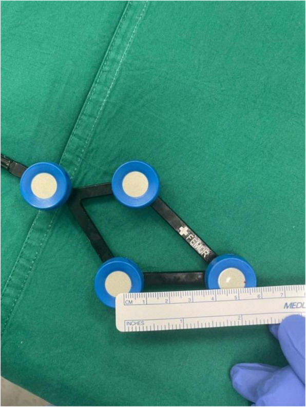
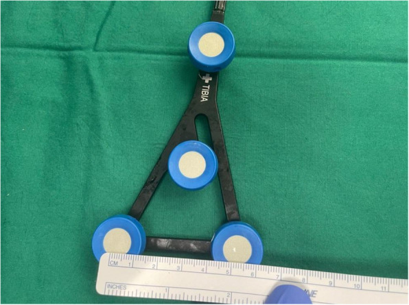
Introduction: Major robotic systems for total knee replacements necessitate the use of array pins in the tibia and femur. These extra-incisional pins are placed away from the primary incision and may be associated with soft tissue complications and peri-prosthetic fractures. There is currently no standardized, reproducible method for reliably placing pins in the femur and tibia metaphyses. We have developed an all-intra-incisional pin method within the primary incision. This paper aims to describe our technique, analyse the proximity of the pins to the implants, and study complications arising from both techniques.
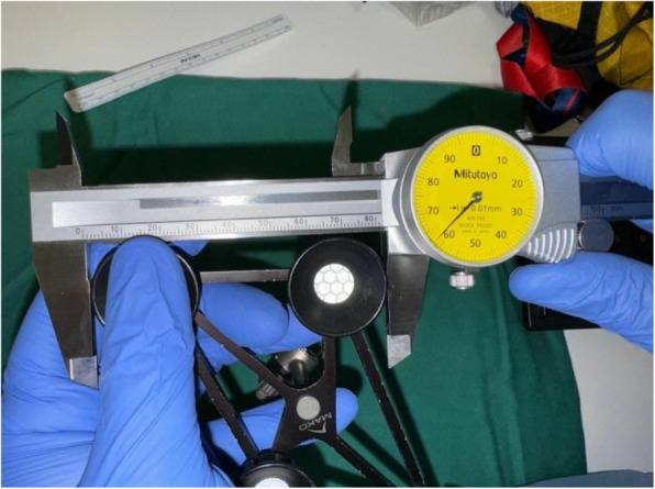
Methods: A total of 102 robotic-assisted total knee arthroplasties were performed using the ROSA, MAKO, and CORI systems. Patient charts were reviewed for their age, gender, body mass index, and ethnicity. Post-operative day zero radiographs of the operated knee were used for measurements in anteroposterior and lateral views, with X-ray magnifications taken into consideration.
Results: Our study demonstrates that intra-incisional pins can be placed 6.52 times closer to the tibial implant compared to extra-incisional pins on the anteroposterior X-ray view radiographs, with no observed significant difference between the complication rates. In anteroposterior view, it allows placement of tibia pins within 8.99 ± 1.21 mm (95% CI: 7.78, 10.2) of the tibial implant, within 5.93 ± 1.29 mm (95% CI: 4.64, 7.22) of the tibia-reamed-surface, and placement of the femoral pins within 6.01 ± 1.37 mm (95% CI: 4.64, 7.37) of the femoral implant. In the lateral X-ray view, it enables the placement of tibial pins within 9.40 ± 1.43 mm (95% CI: 7.97, 10.8) of the implant. Univariate analysis reveals that our technique and pin-distance from the implants are not influenced by patient demographics.
Conclusion: Our study has demonstrated that our technique is precise, not affected by patients' demographics, and eliminates the need for pin repositioning, potentially reducing the incidence of pin-site complications.

 求助内容:
求助内容: 应助结果提醒方式:


