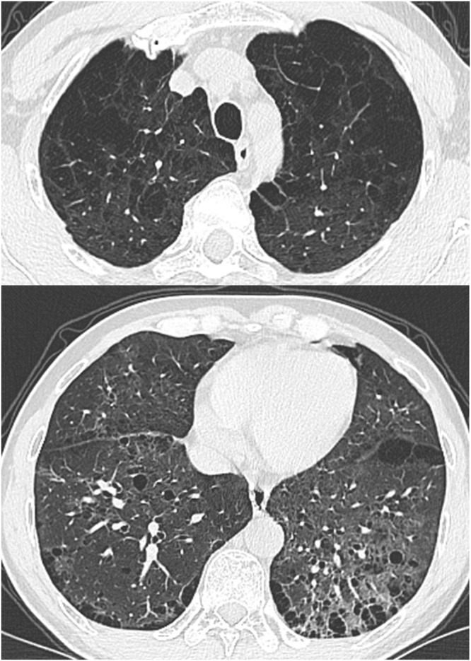
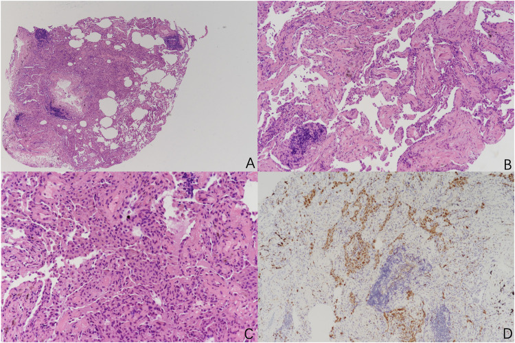
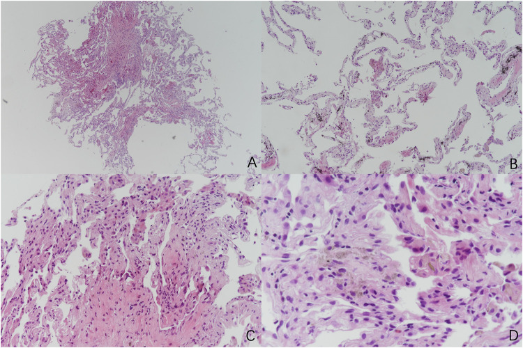
{"title":"The Dual Threat Combined Pulmonary Fibrosis and Emphysema (CPFE): Two Cases.","authors":"Shan Peng, Fanqing Meng, Cheng Wu, Mengshu Cao, Wei Li, Xiaoyan Xin, Anning Feng","doi":"10.2147/COPD.S523597","DOIUrl":null,"url":null,"abstract":"<p><p>Emphysema is common in fibrotic interstitial lung diseases, and its combination with pulmonary fibrosis is known as \"Combined Pulmonary Fibrosis and Emphysema (CPFE) syndrome\". The diagnosis of CPFE significantly impacts treatment strategies and prognosis. In this article, we report the clinical, imaging, and especially the pathological features of two CPFE patients. <b>Case 1</b>: A 51-year-old male patient with a history of smoking. CT scans revealed interstitial lung disease combined with pulmonary bullae. Pathology showed extensive deposition of mononuclear cells in the alveolar spaces, with some cells phagocytosing pigment. Mild fibrous tissue hyperplasia was present in the lung interstitium, along with chronic inflammation and lymphoid nodule formation. The histological findings were consistent with desquamative interstitial pneumonia (DIP), and the clinical, imaging, and pathological correlation confirmed a diagnosis of CPFE. <b>Case 2</b>: A 58-year-old male, a driver with a history of dust exposure and smoking, was admitted due to chest tightness and a cough for 2 years. Chest CT revealed interstitial changes, emphysema, and bullae in both lungs. Histopathology showed fibrous widening of alveolar septa, mild chronic inflammation, and dust cell deposition, along with emphysematous changes and bulla formation, consistent with CPFE. The purpose of this report is to increase pathologists' awareness of this complex disease and emphasize the importance of multidisciplinary cooperation in the diagnosis and treatment of CPFE. Furthermore, this article encourages further research into CPFE.</p>","PeriodicalId":48818,"journal":{"name":"International Journal of Chronic Obstructive Pulmonary Disease","volume":"20 ","pages":"2885-2891"},"PeriodicalIF":3.1000,"publicationDate":"2025-08-15","publicationTypes":"Journal Article","fieldsOfStudy":null,"isOpenAccess":false,"openAccessPdf":"https://www.ncbi.nlm.nih.gov/pmc/articles/PMC12364000/pdf/","citationCount":"0","resultStr":null,"platform":"Semanticscholar","paperid":null,"PeriodicalName":"International Journal of Chronic Obstructive Pulmonary Disease","FirstCategoryId":"3","ListUrlMain":"https://doi.org/10.2147/COPD.S523597","RegionNum":3,"RegionCategory":"医学","ArticlePicture":[],"TitleCN":null,"AbstractTextCN":null,"PMCID":null,"EPubDate":"2025/1/1 0:00:00","PubModel":"eCollection","JCR":"Q2","JCRName":"RESPIRATORY SYSTEM","Score":null,"Total":0}
引用次数: 0
Abstract
Emphysema is common in fibrotic interstitial lung diseases, and its combination with pulmonary fibrosis is known as "Combined Pulmonary Fibrosis and Emphysema (CPFE) syndrome". The diagnosis of CPFE significantly impacts treatment strategies and prognosis. In this article, we report the clinical, imaging, and especially the pathological features of two CPFE patients. Case 1: A 51-year-old male patient with a history of smoking. CT scans revealed interstitial lung disease combined with pulmonary bullae. Pathology showed extensive deposition of mononuclear cells in the alveolar spaces, with some cells phagocytosing pigment. Mild fibrous tissue hyperplasia was present in the lung interstitium, along with chronic inflammation and lymphoid nodule formation. The histological findings were consistent with desquamative interstitial pneumonia (DIP), and the clinical, imaging, and pathological correlation confirmed a diagnosis of CPFE. Case 2: A 58-year-old male, a driver with a history of dust exposure and smoking, was admitted due to chest tightness and a cough for 2 years. Chest CT revealed interstitial changes, emphysema, and bullae in both lungs. Histopathology showed fibrous widening of alveolar septa, mild chronic inflammation, and dust cell deposition, along with emphysematous changes and bulla formation, consistent with CPFE. The purpose of this report is to increase pathologists' awareness of this complex disease and emphasize the importance of multidisciplinary cooperation in the diagnosis and treatment of CPFE. Furthermore, this article encourages further research into CPFE.
期刊介绍:
An international, peer-reviewed journal of therapeutics and pharmacology focusing on concise rapid reporting of clinical studies and reviews in COPD. Special focus will be given to the pathophysiological processes underlying the disease, intervention programs, patient focused education, and self management protocols. This journal is directed at specialists and healthcare professionals
 求助内容:
求助内容: 应助结果提醒方式:


