{"title":"Unilateral Vibratory Stimulation Inhibits Contralateral Spinal Anterior Horn Cells in Homonymous Muscles for the First 75 Seconds.","authors":"Kenta Kunoh, Takahiro Takenaka, Daisuke Kimura, Toshiaki Suzuki","doi":"10.5535/arm.240107","DOIUrl":null,"url":null,"abstract":"<p><strong>Objective: </strong>To investigate muscle tone changes over time in contralateral homonymous muscles when unilateral muscles are stimulated, using F-wave measurements, we examined whether vibratory stimulation on the contralateral homonymous muscle of the affected side may reduce spasticity, whose optimal duration remains unclear.</p><p><strong>Methods: </strong>Vibratory stimulation was applied to the right hand of healthy adults, using parameters of 80 Hz frequency, 0.4 mm amplitude, 400 g load, and 195 seconds of duration on the abductor digiti minimi muscle. F-wave was measured in the left hand before stimulation, at seven intervals during stimulation, and immediately after.</p><p><strong>Results: </strong>The F/M amplitude ratio decreased immediately at stimulation onset, at 30 seconds, and at 60 seconds compared to baseline. A least-squares analysis revealed a negative slope from baseline to 60 seconds (f(x)=-0.11x+1.12), while the slope became positive after 90 seconds, continuing after stimulation ended (f(x)=0.04x+0.82).</p><p><strong>Conclusion: </strong>Unilateral vibratory stimulation may decrease excitability in the spinal anterior horn cells of the contralateral homonymous muscle for up to 75 seconds post-stimulation, suggesting a potential mechanism for spasticity management.</p>","PeriodicalId":47738,"journal":{"name":"Annals of Rehabilitation Medicine-ARM","volume":"49 4","pages":"226-233"},"PeriodicalIF":2.9000,"publicationDate":"2025-08-01","publicationTypes":"Journal Article","fieldsOfStudy":null,"isOpenAccess":false,"openAccessPdf":"https://www.ncbi.nlm.nih.gov/pmc/articles/PMC12411864/pdf/","citationCount":"0","resultStr":null,"platform":"Semanticscholar","paperid":null,"PeriodicalName":"Annals of Rehabilitation Medicine-ARM","FirstCategoryId":"1085","ListUrlMain":"https://doi.org/10.5535/arm.240107","RegionNum":0,"RegionCategory":null,"ArticlePicture":[],"TitleCN":null,"AbstractTextCN":null,"PMCID":null,"EPubDate":"2025/8/22 0:00:00","PubModel":"Epub","JCR":"Q1","JCRName":"REHABILITATION","Score":null,"Total":0}
引用次数: 0
Abstract
Objective: To investigate muscle tone changes over time in contralateral homonymous muscles when unilateral muscles are stimulated, using F-wave measurements, we examined whether vibratory stimulation on the contralateral homonymous muscle of the affected side may reduce spasticity, whose optimal duration remains unclear.
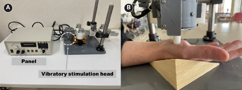
Methods: Vibratory stimulation was applied to the right hand of healthy adults, using parameters of 80 Hz frequency, 0.4 mm amplitude, 400 g load, and 195 seconds of duration on the abductor digiti minimi muscle. F-wave was measured in the left hand before stimulation, at seven intervals during stimulation, and immediately after.
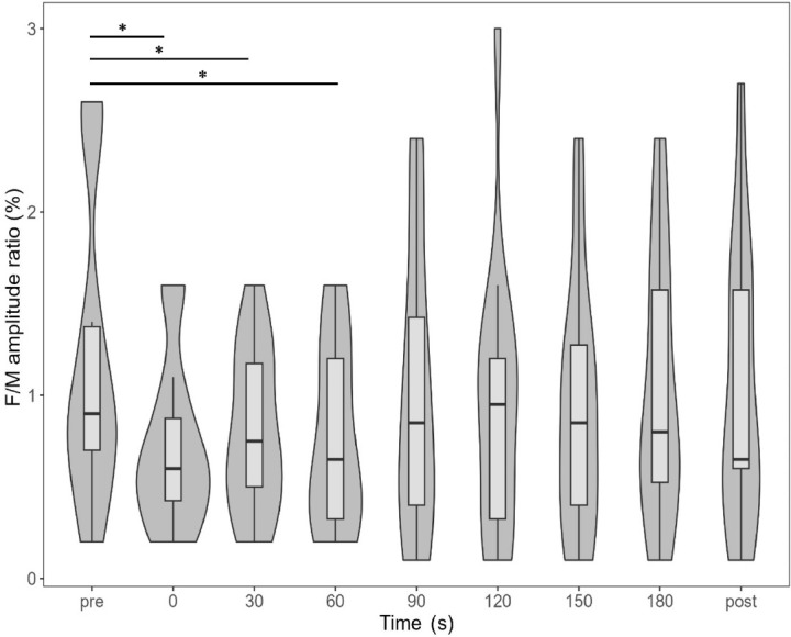
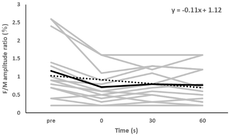
Results: The F/M amplitude ratio decreased immediately at stimulation onset, at 30 seconds, and at 60 seconds compared to baseline. A least-squares analysis revealed a negative slope from baseline to 60 seconds (f(x)=-0.11x+1.12), while the slope became positive after 90 seconds, continuing after stimulation ended (f(x)=0.04x+0.82).
Conclusion: Unilateral vibratory stimulation may decrease excitability in the spinal anterior horn cells of the contralateral homonymous muscle for up to 75 seconds post-stimulation, suggesting a potential mechanism for spasticity management.

 求助内容:
求助内容: 应助结果提醒方式:


