{"title":"Airway Foreign Body-Leave Nothing Behind.","authors":"Jeffrey Ng, Christopher Thong, Kay Choong See","doi":"10.1002/rcr2.70332","DOIUrl":null,"url":null,"abstract":"<p><p>A middle-aged male presented with chronic cough, shortness of breath and fever. In the presence of wheeze and mild right-sided opacities on chest x-ray, he was treated for infective exacerbation of asthma with nebulised bronchodilators, antibiotics and steroids. Due to persistent wheeze, a contrasted computed tomography (CT) scan was done and revealed the presence of two endobronchial linear foreign body (FB) opacities, the second located more distally than the first. A rigid bronchoscopy-based technique under general anaesthesia was used to remove the first FB, and flexible bronchoscopy via endotracheal tube was used for the second FB in the same session. This case illustrates the tenets of airway FB diagnosis and management. The differential diagnosis of radiolucent airway FB needs to be considered in patients with chronic symptoms. Rigid and flexible bronchoscopy techniques are complementary. Peripheral lesions are more accessible by flexible bronchoscopy. Measures to ensure complete removal of FBs must be consistently incorporated into routine practice.</p>","PeriodicalId":45846,"journal":{"name":"Respirology Case Reports","volume":"13 8","pages":"e70332"},"PeriodicalIF":0.8000,"publicationDate":"2025-08-26","publicationTypes":"Journal Article","fieldsOfStudy":null,"isOpenAccess":false,"openAccessPdf":"https://www.ncbi.nlm.nih.gov/pmc/articles/PMC12379834/pdf/","citationCount":"0","resultStr":null,"platform":"Semanticscholar","paperid":null,"PeriodicalName":"Respirology Case Reports","FirstCategoryId":"1085","ListUrlMain":"https://doi.org/10.1002/rcr2.70332","RegionNum":0,"RegionCategory":null,"ArticlePicture":[],"TitleCN":null,"AbstractTextCN":null,"PMCID":null,"EPubDate":"2025/8/1 0:00:00","PubModel":"eCollection","JCR":"Q4","JCRName":"RESPIRATORY SYSTEM","Score":null,"Total":0}
引用次数: 0
Abstract
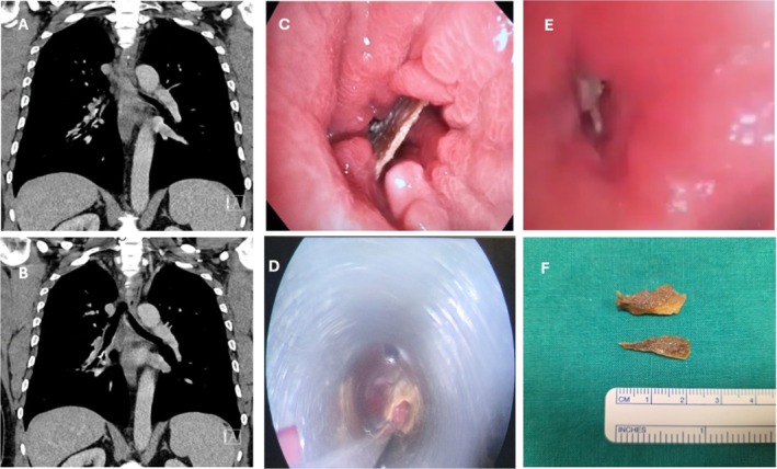
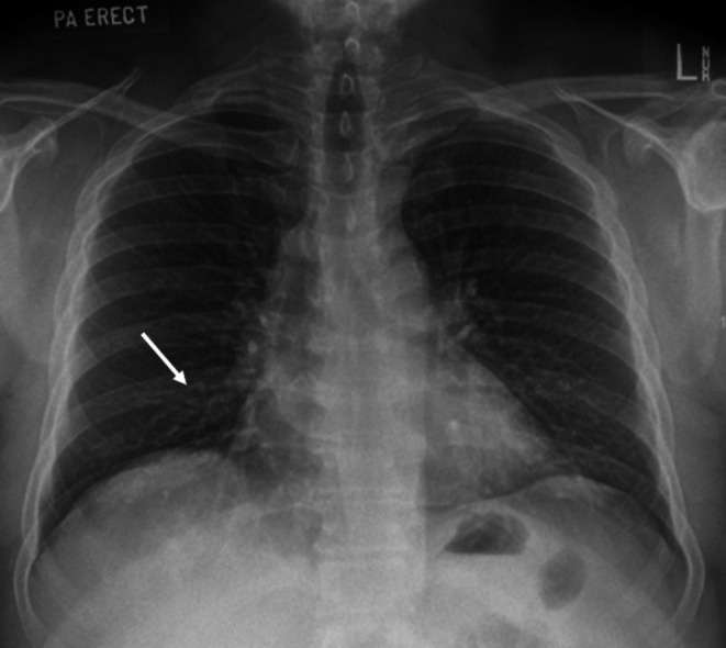
A middle-aged male presented with chronic cough, shortness of breath and fever. In the presence of wheeze and mild right-sided opacities on chest x-ray, he was treated for infective exacerbation of asthma with nebulised bronchodilators, antibiotics and steroids. Due to persistent wheeze, a contrasted computed tomography (CT) scan was done and revealed the presence of two endobronchial linear foreign body (FB) opacities, the second located more distally than the first. A rigid bronchoscopy-based technique under general anaesthesia was used to remove the first FB, and flexible bronchoscopy via endotracheal tube was used for the second FB in the same session. This case illustrates the tenets of airway FB diagnosis and management. The differential diagnosis of radiolucent airway FB needs to be considered in patients with chronic symptoms. Rigid and flexible bronchoscopy techniques are complementary. Peripheral lesions are more accessible by flexible bronchoscopy. Measures to ensure complete removal of FBs must be consistently incorporated into routine practice.
期刊介绍:
Respirology Case Reports is an open-access online journal dedicated to the publication of original clinical case reports, case series, clinical images and clinical videos in all fields of respiratory medicine. The Journal encourages the international exchange between clinicians and researchers of experiences in diagnosing and treating uncommon diseases or diseases with unusual presentations. All manuscripts are peer-reviewed through a streamlined process that aims at providing a rapid turnaround time from submission to publication.


 求助内容:
求助内容: 应助结果提醒方式:


