{"title":"Percentile Distribution of Habitual-Correction Visual Acuity in a Sample of 1500 Children Aged 5 to 15 Years in Italy.","authors":"Alessio Facchin, Marilena Mazzilli, Silvio Maffioletti","doi":"10.3390/pediatric17040085","DOIUrl":null,"url":null,"abstract":"<p><p><b>Background:</b> Early identification of visual disorders in children is essential to prevent long-term visual impairment and support academic development. Despite the recognized importance of visual screenings, no universal consensus exists on which visual parameters or threshold values should be used, particularly for measuring visual acuity (VA) in pediatric populations. <b>Objectives:</b> This study aimed to develop age-related percentile norms for VA using LEA symbol charts. <b>Methods</b>: A sample of Italian schoolchildren aged 5 to 15 years (<i>n</i> = 1510) participated in the study. Data were collected retrospectively from school-based vision screenings conducted across 12 schools in the Lombardy and Piedmont regions from 2010 to 2019. Monocular and binocular VA were measured at 3 m using a standardized LEA symbol chart, and values were scored letter-by-letter on a LogMAR scale. Smoothed percentile curves were derived using Box-Cox, Cole, and Green distribution modeling and regression analysis. <b>Results:</b> The results showed a non-linear improvement in VA with age. Compared to prior studies, LEA symbols yielded slightly lower VA scores, reinforcing the need for chart-specific norms. The 50th percentile VA improved from approximately +0.07 LogMAR at age 6 to about -0.09 LogMAR at age 15. <b>Conclusions</b>: These findings highlight the importance of age-specific, chart-specific, and statistically robust reference data for VA screening in children. The derived percentile tables offer a more sensitive tool than fixed cut-offs for identifying visual anomalies and tailoring clinical interventions. This work contributes to standardizing pediatric VA screening practices and improving early detection of visual deficits.</p>","PeriodicalId":45251,"journal":{"name":"Pediatric Reports","volume":"17 4","pages":""},"PeriodicalIF":1.4000,"publicationDate":"2025-08-11","publicationTypes":"Journal Article","fieldsOfStudy":null,"isOpenAccess":false,"openAccessPdf":"https://www.ncbi.nlm.nih.gov/pmc/articles/PMC12389372/pdf/","citationCount":"0","resultStr":null,"platform":"Semanticscholar","paperid":null,"PeriodicalName":"Pediatric Reports","FirstCategoryId":"1085","ListUrlMain":"https://doi.org/10.3390/pediatric17040085","RegionNum":0,"RegionCategory":null,"ArticlePicture":[],"TitleCN":null,"AbstractTextCN":null,"PMCID":null,"EPubDate":"","PubModel":"","JCR":"Q3","JCRName":"PEDIATRICS","Score":null,"Total":0}
引用次数: 0
Abstract
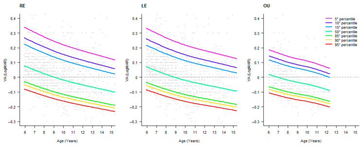
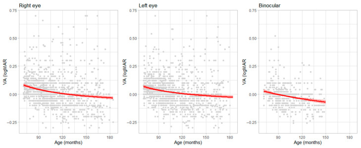
Background: Early identification of visual disorders in children is essential to prevent long-term visual impairment and support academic development. Despite the recognized importance of visual screenings, no universal consensus exists on which visual parameters or threshold values should be used, particularly for measuring visual acuity (VA) in pediatric populations. Objectives: This study aimed to develop age-related percentile norms for VA using LEA symbol charts. Methods: A sample of Italian schoolchildren aged 5 to 15 years (n = 1510) participated in the study. Data were collected retrospectively from school-based vision screenings conducted across 12 schools in the Lombardy and Piedmont regions from 2010 to 2019. Monocular and binocular VA were measured at 3 m using a standardized LEA symbol chart, and values were scored letter-by-letter on a LogMAR scale. Smoothed percentile curves were derived using Box-Cox, Cole, and Green distribution modeling and regression analysis. Results: The results showed a non-linear improvement in VA with age. Compared to prior studies, LEA symbols yielded slightly lower VA scores, reinforcing the need for chart-specific norms. The 50th percentile VA improved from approximately +0.07 LogMAR at age 6 to about -0.09 LogMAR at age 15. Conclusions: These findings highlight the importance of age-specific, chart-specific, and statistically robust reference data for VA screening in children. The derived percentile tables offer a more sensitive tool than fixed cut-offs for identifying visual anomalies and tailoring clinical interventions. This work contributes to standardizing pediatric VA screening practices and improving early detection of visual deficits.

 求助内容:
求助内容: 应助结果提醒方式:


