Lili Miles, Caroline Baughn, Gleidson Messias Silva, Dorothea L Douglas, Lei Shao
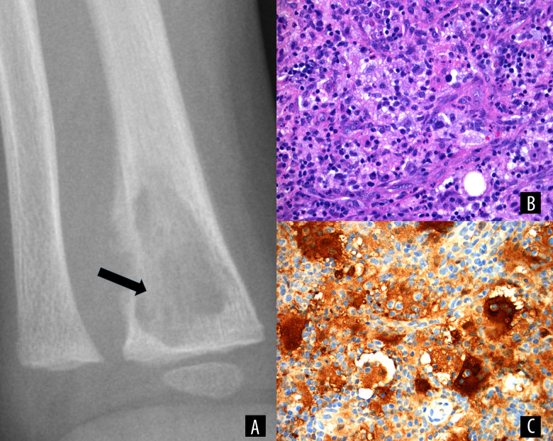
{"title":"Extranodal Rosai-Dorfman Disease in a Pediatric Patient: A Case Report.","authors":"Lili Miles, Caroline Baughn, Gleidson Messias Silva, Dorothea L Douglas, Lei Shao","doi":"10.12659/AJCR.948533","DOIUrl":null,"url":null,"abstract":"<p><p>BACKGROUND Sinus histiocytosis with massive lymphadenopathy (also known as Rosai-Dorfman disease [RDD]), was originally recognized in 1969 by Rosai and Dorfman. RDD is characterized by the accumulation of activated histiocytes in various tissues and organs, but most commonly in lymph nodes. RDD is subclassified into 2 forms. The more common form, nodal RDD, and the rare form, extranodal RDD, which is based on the presence of extranodal tissue involvement. Most extranodal RDD cases also involve lymph nodes. Primary, exclusively extranodal RDD is rare, and is exceedingly uncommon in young pediatric patients. These cases have rarely been reported in the medical literature. CASE REPORT This 16-month-old previously healthy boy presented with right-forearm pain, following a right-wrist injury. Imaging studies revealed an aggressive lesion of the distal radius with the presence of an adjacent soft-tissue component. The core-needle tissue biopsy suggested osteomyelitis. Because the patient was not responsive to antibiotics, he underwent curettage with washout for the management of presumed osteomyelitis. RDD was diagnosed based on the curettage material. Subsequent imaging studies confirmed the isolated right-radius bone lesion without any other organ or tissue involvement. He received observation management with surveillance imaging every 6 months. Thirty-six months later, he was healthy, without pain, and had normal right-wrist function. CONCLUSIONS Primary osseous RDD in children is exceedingly rare and can cause diagnostic challenges. Our case serves as a reminder that despite the diagnostic challenge, keeping RDD in the differential diagnosis, especially in young patients, can avoid misdiagnosis and mismanagement.</p>","PeriodicalId":39064,"journal":{"name":"American Journal of Case Reports","volume":"26 ","pages":"e948533"},"PeriodicalIF":0.7000,"publicationDate":"2025-08-27","publicationTypes":"Journal Article","fieldsOfStudy":null,"isOpenAccess":false,"openAccessPdf":"https://www.ncbi.nlm.nih.gov/pmc/articles/PMC12400872/pdf/","citationCount":"0","resultStr":null,"platform":"Semanticscholar","paperid":null,"PeriodicalName":"American Journal of Case Reports","FirstCategoryId":"1085","ListUrlMain":"https://doi.org/10.12659/AJCR.948533","RegionNum":0,"RegionCategory":null,"ArticlePicture":[],"TitleCN":null,"AbstractTextCN":null,"PMCID":null,"EPubDate":"","PubModel":"","JCR":"Q3","JCRName":"MEDICINE, GENERAL & INTERNAL","Score":null,"Total":0}
引用次数: 0
Abstract
BACKGROUND Sinus histiocytosis with massive lymphadenopathy (also known as Rosai-Dorfman disease [RDD]), was originally recognized in 1969 by Rosai and Dorfman. RDD is characterized by the accumulation of activated histiocytes in various tissues and organs, but most commonly in lymph nodes. RDD is subclassified into 2 forms. The more common form, nodal RDD, and the rare form, extranodal RDD, which is based on the presence of extranodal tissue involvement. Most extranodal RDD cases also involve lymph nodes. Primary, exclusively extranodal RDD is rare, and is exceedingly uncommon in young pediatric patients. These cases have rarely been reported in the medical literature. CASE REPORT This 16-month-old previously healthy boy presented with right-forearm pain, following a right-wrist injury. Imaging studies revealed an aggressive lesion of the distal radius with the presence of an adjacent soft-tissue component. The core-needle tissue biopsy suggested osteomyelitis. Because the patient was not responsive to antibiotics, he underwent curettage with washout for the management of presumed osteomyelitis. RDD was diagnosed based on the curettage material. Subsequent imaging studies confirmed the isolated right-radius bone lesion without any other organ or tissue involvement. He received observation management with surveillance imaging every 6 months. Thirty-six months later, he was healthy, without pain, and had normal right-wrist function. CONCLUSIONS Primary osseous RDD in children is exceedingly rare and can cause diagnostic challenges. Our case serves as a reminder that despite the diagnostic challenge, keeping RDD in the differential diagnosis, especially in young patients, can avoid misdiagnosis and mismanagement.
期刊介绍:
American Journal of Case Reports is an international, peer-reviewed scientific journal that publishes single and series case reports in all medical fields. American Journal of Case Reports is issued on a continuous basis as a primary electronic journal. Print copies of a single article or a set of articles can be ordered on demand.

 求助内容:
求助内容: 应助结果提醒方式:


