{"title":"An Older Patient with a Symptomatic Arachnoid Cyst in the Velum Interpositum: Considerations of Functional Neuroanatomy.","authors":"Shunsuke Fujitsuku, Sadahiro Nomura, Hirokazu Sadahiro, Masami Osaki, Hideyuki Ishihara","doi":"10.1055/a-2678-8527","DOIUrl":null,"url":null,"abstract":"<p><p>We report a patient with an arachnoid cyst in the velum interpositum (VI) and discuss the mechanism of the symptoms based on functional neuroanatomy. A 68-year-old woman presented with difficulty in doing housekeeping and with route-finding disorientation in known locations. Her performance intelligence quotient (PIQ) score was 68, significantly lower than her verbal intelligence quotient (IQ) of 103. Significantly low scores were obtained for the picture arrangement, picture completion, and symbol search tasks (4, 1, and 5, respectively) in the PIQ subtests. Her copies of the interlocking pentagons and cube designs were distorted, indicating visual-spatial construction apraxia. However, verbal IQ, working memory, urination control, ideational and ideomotor function, and dressing were intact. Magnetic resonance imaging revealed a cystic enlargement of the VI. Neuroendoscopic cyst fenestration to the lateral ventricles contributed to a decrease in the volume of the cyst. Postoperatively, her PIQ improved to 94. Her scores on the picture arrangement, picture completion, and symbol search tests increased to 7, 7, and 11 points, respectively. The pentagons and cube designs were copied correctly. An arachnoid cyst in VI is known to present with cognitive dysfunction. In our patient, symptoms were limited to the constructional apraxia and route-finding disorientation owing to the disturbance in the biparietal connections and posterior cingulate gyrus, respectively. The intramantle pressure gradient created by the characteristic cone-shaped cyst may have caused the selective dysfunctions. Namely, the impairment in the deep parietal region was more severe than on the frontal lobes or superficial parietal lobes.</p>","PeriodicalId":44256,"journal":{"name":"Journal of Neurological Surgery Reports","volume":"86 3","pages":"e180-e184"},"PeriodicalIF":0.7000,"publicationDate":"2025-08-21","publicationTypes":"Journal Article","fieldsOfStudy":null,"isOpenAccess":false,"openAccessPdf":"https://www.ncbi.nlm.nih.gov/pmc/articles/PMC12370392/pdf/","citationCount":"0","resultStr":null,"platform":"Semanticscholar","paperid":null,"PeriodicalName":"Journal of Neurological Surgery Reports","FirstCategoryId":"1085","ListUrlMain":"https://doi.org/10.1055/a-2678-8527","RegionNum":0,"RegionCategory":null,"ArticlePicture":[],"TitleCN":null,"AbstractTextCN":null,"PMCID":null,"EPubDate":"2025/7/1 0:00:00","PubModel":"eCollection","JCR":"Q4","JCRName":"CLINICAL NEUROLOGY","Score":null,"Total":0}
引用次数: 0
Abstract
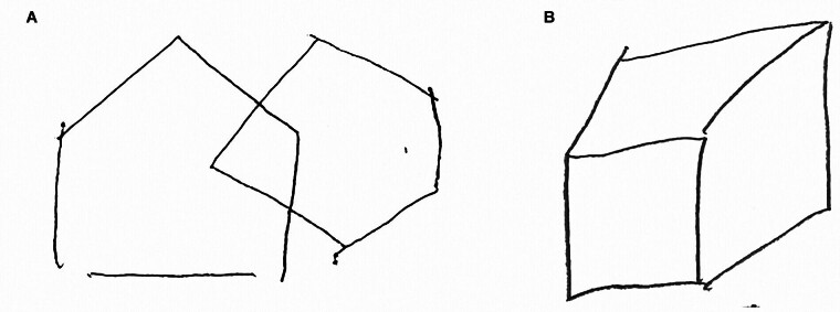
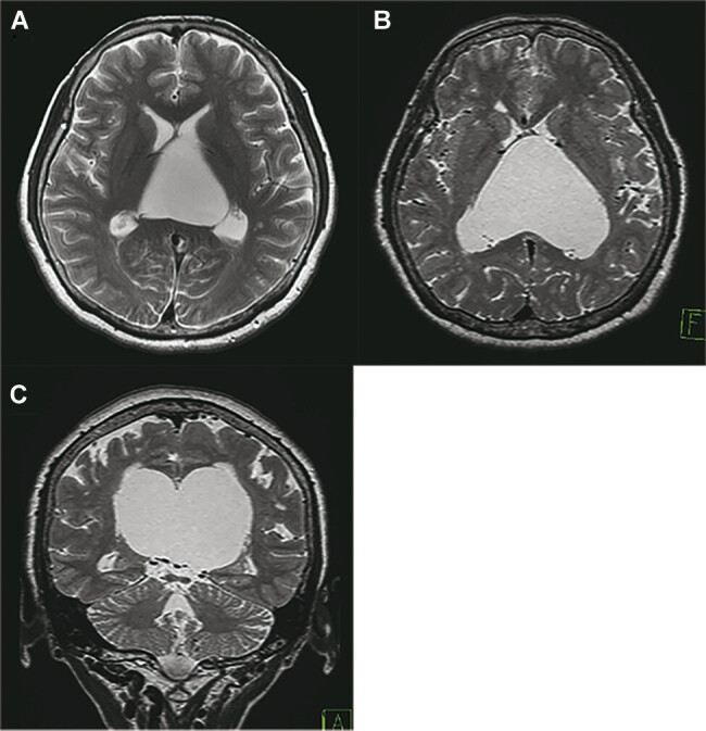
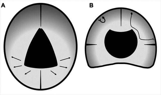
We report a patient with an arachnoid cyst in the velum interpositum (VI) and discuss the mechanism of the symptoms based on functional neuroanatomy. A 68-year-old woman presented with difficulty in doing housekeeping and with route-finding disorientation in known locations. Her performance intelligence quotient (PIQ) score was 68, significantly lower than her verbal intelligence quotient (IQ) of 103. Significantly low scores were obtained for the picture arrangement, picture completion, and symbol search tasks (4, 1, and 5, respectively) in the PIQ subtests. Her copies of the interlocking pentagons and cube designs were distorted, indicating visual-spatial construction apraxia. However, verbal IQ, working memory, urination control, ideational and ideomotor function, and dressing were intact. Magnetic resonance imaging revealed a cystic enlargement of the VI. Neuroendoscopic cyst fenestration to the lateral ventricles contributed to a decrease in the volume of the cyst. Postoperatively, her PIQ improved to 94. Her scores on the picture arrangement, picture completion, and symbol search tests increased to 7, 7, and 11 points, respectively. The pentagons and cube designs were copied correctly. An arachnoid cyst in VI is known to present with cognitive dysfunction. In our patient, symptoms were limited to the constructional apraxia and route-finding disorientation owing to the disturbance in the biparietal connections and posterior cingulate gyrus, respectively. The intramantle pressure gradient created by the characteristic cone-shaped cyst may have caused the selective dysfunctions. Namely, the impairment in the deep parietal region was more severe than on the frontal lobes or superficial parietal lobes.

 求助内容:
求助内容: 应助结果提醒方式:


