Mohammad Al-Hurani, Yousef Al-Hadrab, Khaleel Raslan Ayoub, Amjad Al-Salhi, Hala Sadi Al-Sarsour, Gregor Jan Kocher
{"title":"Thoracoscopic Approach for Treating a Primary Hydatid Cyst in the Thymus in a Teenager: A Case Report.","authors":"Mohammad Al-Hurani, Yousef Al-Hadrab, Khaleel Raslan Ayoub, Amjad Al-Salhi, Hala Sadi Al-Sarsour, Gregor Jan Kocher","doi":"10.12659/AJCR.948600","DOIUrl":null,"url":null,"abstract":"<p><p>BACKGROUND Hydatid disease remains a major clinical concern, particularly in regions where it is endemic. This parasitic infection is caused by Echinococcus species. The liver and lungs are the most affected organs. Although the lungs are the most commonly affected intrathoracic organ, extrapulmonary intrathoracic hydatid disease is uncommon. However, primary mediastinal hydatid disease is a rare entity, and a primary hydatid cyst in the thymus is extremely rare. Although video-assisted thoracoscopic surgery (VATS) plays an important role in the field of thoracic surgery, its role in treating hydatid disease in the chest is not well established. This report describes a case of 17-year-old male presenting with primary hydatid cyst of the thymus treated thoracoscopically. CASE REPORT A 17-year-old male presented to our clinic with chest tightness and shortness of breath of 3 months duration. A well-defined opacity was identified on chest radiography. Subsequent computed tomography (CT) revealed a large, well-defined cystic lesion in the left hemithorax, measuring 6.5×7×11 cm. He was later scheduled for VATS to resect the cyst, which was found to be in continuity with the thymic gland. An en bloc thymectomy was performed to ensure complete peri-cystectomy. CONCLUSIONS This case is unique not only because it describes a rare location of hydatid disease, but also due to the technique of resection that was used. Based on our literature review, this is among the earliest reported cases of a hydatid cyst in this location resected via thoracoscopy. Furthermore, compared with thoracotomy, VATS offers faster recovery and reduced postoperative pain, and its application in similar cases should be further explored.</p>","PeriodicalId":39064,"journal":{"name":"American Journal of Case Reports","volume":"26 ","pages":"e948600"},"PeriodicalIF":0.7000,"publicationDate":"2025-08-22","publicationTypes":"Journal Article","fieldsOfStudy":null,"isOpenAccess":false,"openAccessPdf":"https://www.ncbi.nlm.nih.gov/pmc/articles/PMC12379743/pdf/","citationCount":"0","resultStr":null,"platform":"Semanticscholar","paperid":null,"PeriodicalName":"American Journal of Case Reports","FirstCategoryId":"1085","ListUrlMain":"https://doi.org/10.12659/AJCR.948600","RegionNum":0,"RegionCategory":null,"ArticlePicture":[],"TitleCN":null,"AbstractTextCN":null,"PMCID":null,"EPubDate":"","PubModel":"","JCR":"Q3","JCRName":"MEDICINE, GENERAL & INTERNAL","Score":null,"Total":0}
引用次数: 0
Abstract
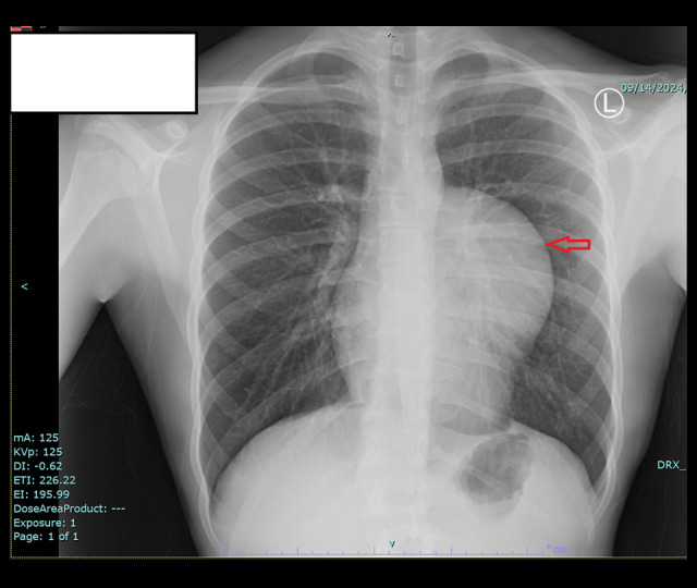
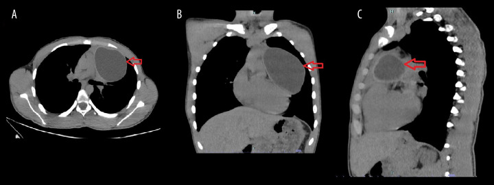
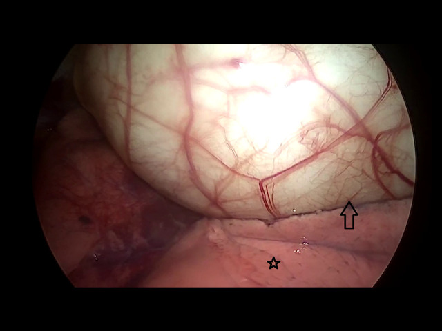
BACKGROUND Hydatid disease remains a major clinical concern, particularly in regions where it is endemic. This parasitic infection is caused by Echinococcus species. The liver and lungs are the most affected organs. Although the lungs are the most commonly affected intrathoracic organ, extrapulmonary intrathoracic hydatid disease is uncommon. However, primary mediastinal hydatid disease is a rare entity, and a primary hydatid cyst in the thymus is extremely rare. Although video-assisted thoracoscopic surgery (VATS) plays an important role in the field of thoracic surgery, its role in treating hydatid disease in the chest is not well established. This report describes a case of 17-year-old male presenting with primary hydatid cyst of the thymus treated thoracoscopically. CASE REPORT A 17-year-old male presented to our clinic with chest tightness and shortness of breath of 3 months duration. A well-defined opacity was identified on chest radiography. Subsequent computed tomography (CT) revealed a large, well-defined cystic lesion in the left hemithorax, measuring 6.5×7×11 cm. He was later scheduled for VATS to resect the cyst, which was found to be in continuity with the thymic gland. An en bloc thymectomy was performed to ensure complete peri-cystectomy. CONCLUSIONS This case is unique not only because it describes a rare location of hydatid disease, but also due to the technique of resection that was used. Based on our literature review, this is among the earliest reported cases of a hydatid cyst in this location resected via thoracoscopy. Furthermore, compared with thoracotomy, VATS offers faster recovery and reduced postoperative pain, and its application in similar cases should be further explored.
期刊介绍:
American Journal of Case Reports is an international, peer-reviewed scientific journal that publishes single and series case reports in all medical fields. American Journal of Case Reports is issued on a continuous basis as a primary electronic journal. Print copies of a single article or a set of articles can be ordered on demand.

 求助内容:
求助内容: 应助结果提醒方式:


