{"title":"Rapid Progression of Primary Hepatic Neuroendocrine Carcinoma: A Case Report Demonstrating Drastic Oncological Behavior.","authors":"Rina Kobayashi, Tomohide Hori, Makoto Yamawaki, Shigeki Nakayama, Satoru Umegae, Takao Iwanaga, Ryutaro Nishikawa, Takahiro Shimoyama, Sakurako Suzuki, Shinichiro Atsumi, Hiroshi Hasegawa, Shigehito Nakashima, Kunihiro Higuchi, Kentaro Onishi, Ryotaro Sakaguchi, Shoichi Morita, Haruka Miyao, Saki Aota, Hikaru Ohtani, Takayuki Yamamoto","doi":"10.12659/AJCR.948500","DOIUrl":null,"url":null,"abstract":"<p><p>BACKGROUND Primary hepatic neuroendocrine neoplasms (PHNENs), including primary hepatic neuroendocrine carcinoma (PHNEC), are extremely rare. PHNENs typically exhibit slow growth, although mixed neuroendocrine-non-neuroendocrine neoplasms have poor prognoses. PHNENs are also challenging to diagnose. CASE REPORT A 73-year-old man underwent plain computed tomography (CT), which incidentally detected a 42-mm solitary hepatic tumor. Serum levels of protein induced by vitamin K absence or antagonist-II (PIVKA-II) were elevated at 138 mAU/mL. Thirteen days later, magnetic resonance imaging (MRI) revealed an enlarged hepatic tumor with tumor thromboses extending into the hepatic and portal veins. No early-phase enhancement was observed. At 18 days, Doppler ultrasound and dynamic CT evaluated the tumor as hypovascular, and a newly swollen solitary lymph node appeared. At 39 days, positron emission tomography (PET)/CT revealed strong uptake in the primary liver tumor and metastatic lymph nodes, with additional distant lymph node metastases emerging. At 49 days, a metastatic cervical lymph node was surgically resected. At 61 days, PHNEC was definitively diagnosed based on histopathological and immunohistochemical assessments. The Ki-67 labeling index was >90%. At 67 days, he was hospitalized to begin chemotherapy, but CT revealed end-stage disease. Palliative treatment was required, and the patient died of cancer 82 days after the initial diagnosis. CONCLUSIONS We have presented a thought-provoking case of PHNEC with rapid oncological progression. To clarify clinical implications (eg, atypical imaging features and diagnostic pitfalls), detailed imaging findings are provided. We anticipate that this case will be informative for clinicians in this field.</p>","PeriodicalId":39064,"journal":{"name":"American Journal of Case Reports","volume":"26 ","pages":"e948500"},"PeriodicalIF":0.7000,"publicationDate":"2025-08-25","publicationTypes":"Journal Article","fieldsOfStudy":null,"isOpenAccess":false,"openAccessPdf":"https://www.ncbi.nlm.nih.gov/pmc/articles/PMC12396091/pdf/","citationCount":"0","resultStr":null,"platform":"Semanticscholar","paperid":null,"PeriodicalName":"American Journal of Case Reports","FirstCategoryId":"1085","ListUrlMain":"https://doi.org/10.12659/AJCR.948500","RegionNum":0,"RegionCategory":null,"ArticlePicture":[],"TitleCN":null,"AbstractTextCN":null,"PMCID":null,"EPubDate":"","PubModel":"","JCR":"Q3","JCRName":"MEDICINE, GENERAL & INTERNAL","Score":null,"Total":0}
引用次数: 0
Abstract
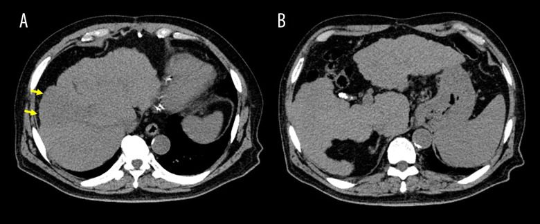
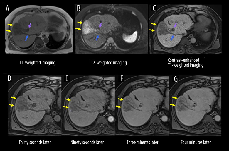
BACKGROUND Primary hepatic neuroendocrine neoplasms (PHNENs), including primary hepatic neuroendocrine carcinoma (PHNEC), are extremely rare. PHNENs typically exhibit slow growth, although mixed neuroendocrine-non-neuroendocrine neoplasms have poor prognoses. PHNENs are also challenging to diagnose. CASE REPORT A 73-year-old man underwent plain computed tomography (CT), which incidentally detected a 42-mm solitary hepatic tumor. Serum levels of protein induced by vitamin K absence or antagonist-II (PIVKA-II) were elevated at 138 mAU/mL. Thirteen days later, magnetic resonance imaging (MRI) revealed an enlarged hepatic tumor with tumor thromboses extending into the hepatic and portal veins. No early-phase enhancement was observed. At 18 days, Doppler ultrasound and dynamic CT evaluated the tumor as hypovascular, and a newly swollen solitary lymph node appeared. At 39 days, positron emission tomography (PET)/CT revealed strong uptake in the primary liver tumor and metastatic lymph nodes, with additional distant lymph node metastases emerging. At 49 days, a metastatic cervical lymph node was surgically resected. At 61 days, PHNEC was definitively diagnosed based on histopathological and immunohistochemical assessments. The Ki-67 labeling index was >90%. At 67 days, he was hospitalized to begin chemotherapy, but CT revealed end-stage disease. Palliative treatment was required, and the patient died of cancer 82 days after the initial diagnosis. CONCLUSIONS We have presented a thought-provoking case of PHNEC with rapid oncological progression. To clarify clinical implications (eg, atypical imaging features and diagnostic pitfalls), detailed imaging findings are provided. We anticipate that this case will be informative for clinicians in this field.
期刊介绍:
American Journal of Case Reports is an international, peer-reviewed scientific journal that publishes single and series case reports in all medical fields. American Journal of Case Reports is issued on a continuous basis as a primary electronic journal. Print copies of a single article or a set of articles can be ordered on demand.

 求助内容:
求助内容: 应助结果提醒方式:


