Sabrina Holguin, Jennifer R Michaels, Michele James
{"title":"Clinical and MRI findings of hemorrhagic infarcts in two cats with positive outcomes.","authors":"Sabrina Holguin, Jennifer R Michaels, Michele James","doi":"10.1177/20551169251360624","DOIUrl":null,"url":null,"abstract":"<p><strong>Case series summary: </strong>A 1.5-year-old male neutered domestic shorthair cat presented with an acute onset of lethargy and difficulty jumping. Prior medical history included a restrictive perimembranous ventricular septal defect and mild mitral regurgitation. Neurologic examination showed left hemiparesis and absent menace in the left eye. MRI revealed a lesion in the right frontal lobe and caudate nucleus with intralesional hemorrhage and restricted diffusion consistent with hemorrhagic infarct. A 6-year-old male neutered domestic shorthair cat presented with three neurologic episodes over 3 months, one consisting of circling to the right, absent menace in the right eye and right-sided postural reaction deficits, and the other two consisting of vestibular signs. MRI revealed a chronic hemorrhagic infarct of the left caudate nucleus. Both cats demonstrated favorable recovery, with cat 2 experiencing a fourth event 9 months after MRI.</p><p><strong>Relevance and novel information: </strong>This case series details the first two cases of feline hemorrhagic infarct with ante-mortem diagnosis, MRI findings and recovery with a good long-term outcome. Hemorrhagic infarcts, uncommonly reported in companion animals, are a subtype of stroke involving hemorrhage resulting from reperfusion or collateral circulation into an ischemic area of brain tissue. This report discusses typical MRI findings in humans, including differentiation from intracerebral hemorrhage, and the potential role of comorbidities on the development and outcome of hemorrhagic infarcts in cats.</p>","PeriodicalId":36588,"journal":{"name":"Journal of Feline Medicine and Surgery Open Reports","volume":"11 2","pages":"20551169251360624"},"PeriodicalIF":0.7000,"publicationDate":"2025-08-26","publicationTypes":"Journal Article","fieldsOfStudy":null,"isOpenAccess":false,"openAccessPdf":"https://www.ncbi.nlm.nih.gov/pmc/articles/PMC12381502/pdf/","citationCount":"0","resultStr":null,"platform":"Semanticscholar","paperid":null,"PeriodicalName":"Journal of Feline Medicine and Surgery Open Reports","FirstCategoryId":"1085","ListUrlMain":"https://doi.org/10.1177/20551169251360624","RegionNum":0,"RegionCategory":null,"ArticlePicture":[],"TitleCN":null,"AbstractTextCN":null,"PMCID":null,"EPubDate":"2025/7/1 0:00:00","PubModel":"eCollection","JCR":"Q3","JCRName":"VETERINARY SCIENCES","Score":null,"Total":0}
引用次数: 0
Abstract
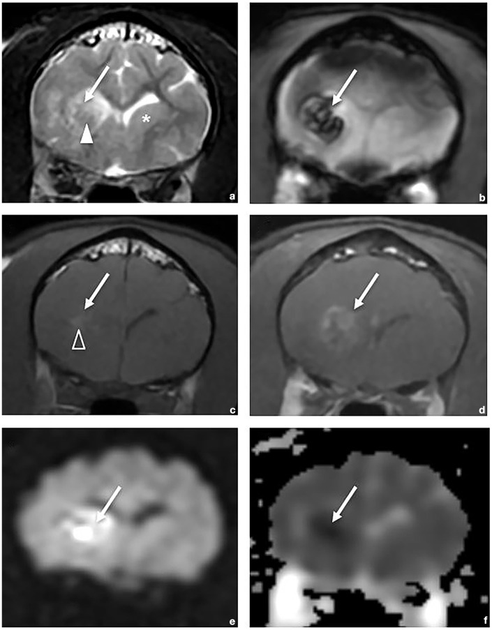
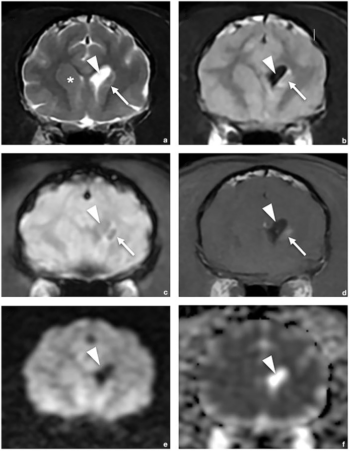
Case series summary: A 1.5-year-old male neutered domestic shorthair cat presented with an acute onset of lethargy and difficulty jumping. Prior medical history included a restrictive perimembranous ventricular septal defect and mild mitral regurgitation. Neurologic examination showed left hemiparesis and absent menace in the left eye. MRI revealed a lesion in the right frontal lobe and caudate nucleus with intralesional hemorrhage and restricted diffusion consistent with hemorrhagic infarct. A 6-year-old male neutered domestic shorthair cat presented with three neurologic episodes over 3 months, one consisting of circling to the right, absent menace in the right eye and right-sided postural reaction deficits, and the other two consisting of vestibular signs. MRI revealed a chronic hemorrhagic infarct of the left caudate nucleus. Both cats demonstrated favorable recovery, with cat 2 experiencing a fourth event 9 months after MRI.
Relevance and novel information: This case series details the first two cases of feline hemorrhagic infarct with ante-mortem diagnosis, MRI findings and recovery with a good long-term outcome. Hemorrhagic infarcts, uncommonly reported in companion animals, are a subtype of stroke involving hemorrhage resulting from reperfusion or collateral circulation into an ischemic area of brain tissue. This report discusses typical MRI findings in humans, including differentiation from intracerebral hemorrhage, and the potential role of comorbidities on the development and outcome of hemorrhagic infarcts in cats.


 求助内容:
求助内容: 应助结果提醒方式:


