Marie Japv Pantangco, Sorcha Costello, Rachel M Basa, Melanie Olive
{"title":"Video-assisted thoracoscopic-guided reduction and external stabilisation of traumatic rib fractures in a cat.","authors":"Marie Japv Pantangco, Sorcha Costello, Rachel M Basa, Melanie Olive","doi":"10.1177/20551169251360620","DOIUrl":null,"url":null,"abstract":"<p><strong>Case summary: </strong>An 11-year-old male neutered domestic longhair cat was presented to an emergency referral hospital after sustaining severe injuries isolated to the thorax after a dog attack. Initial stabilisation included oxygen supplementation, intravenous fluid therapy, point-of-care ultrasound (POCUS), serial blood gas analyses, thoracic and abdominal radiographs, opioid analgesics and broad-spectrum antibiotics. CT revealed multiple internally displaced rib fractures that punctured through the mediastinum and were in intimate proximity to the cranial vena cava and proximal aortic arch. Video-assisted thoracoscopic surgery (VATS) was performed using a 2.7 mm 0° short laparoscope to place a custom-made fibre glass splint via percutaneous suturing to accurately reduce the rib fracture fragments without further damage to the intrathoracic structures. The splint remained in place for 6 weeks. At the 8-week recheck, the cat had no evidence of cardiovascular or respiratory compromise and was able to resume normal activity. A long-term follow-up phone call was conducted at 14 months postoperatively. The owner reported that the patient has continued to do well without any complications.</p><p><strong>Relevance and novel information: </strong>To the authors' knowledge, this is the first report using VATS for the reduction of traumatic rib fractures in a cat. This case highlights the feasibility and success of this method and is a viable option in future clinical cases.</p>","PeriodicalId":36588,"journal":{"name":"Journal of Feline Medicine and Surgery Open Reports","volume":"11 2","pages":"20551169251360620"},"PeriodicalIF":0.7000,"publicationDate":"2025-08-26","publicationTypes":"Journal Article","fieldsOfStudy":null,"isOpenAccess":false,"openAccessPdf":"https://www.ncbi.nlm.nih.gov/pmc/articles/PMC12381480/pdf/","citationCount":"0","resultStr":null,"platform":"Semanticscholar","paperid":null,"PeriodicalName":"Journal of Feline Medicine and Surgery Open Reports","FirstCategoryId":"1085","ListUrlMain":"https://doi.org/10.1177/20551169251360620","RegionNum":0,"RegionCategory":null,"ArticlePicture":[],"TitleCN":null,"AbstractTextCN":null,"PMCID":null,"EPubDate":"2025/7/1 0:00:00","PubModel":"eCollection","JCR":"Q3","JCRName":"VETERINARY SCIENCES","Score":null,"Total":0}
引用次数: 0
Abstract
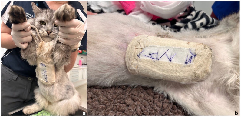
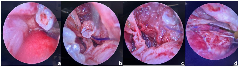
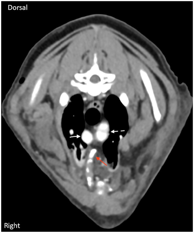
Case summary: An 11-year-old male neutered domestic longhair cat was presented to an emergency referral hospital after sustaining severe injuries isolated to the thorax after a dog attack. Initial stabilisation included oxygen supplementation, intravenous fluid therapy, point-of-care ultrasound (POCUS), serial blood gas analyses, thoracic and abdominal radiographs, opioid analgesics and broad-spectrum antibiotics. CT revealed multiple internally displaced rib fractures that punctured through the mediastinum and were in intimate proximity to the cranial vena cava and proximal aortic arch. Video-assisted thoracoscopic surgery (VATS) was performed using a 2.7 mm 0° short laparoscope to place a custom-made fibre glass splint via percutaneous suturing to accurately reduce the rib fracture fragments without further damage to the intrathoracic structures. The splint remained in place for 6 weeks. At the 8-week recheck, the cat had no evidence of cardiovascular or respiratory compromise and was able to resume normal activity. A long-term follow-up phone call was conducted at 14 months postoperatively. The owner reported that the patient has continued to do well without any complications.
Relevance and novel information: To the authors' knowledge, this is the first report using VATS for the reduction of traumatic rib fractures in a cat. This case highlights the feasibility and success of this method and is a viable option in future clinical cases.
病例总结:一只11岁的雄性绝育家长毛猫在被狗袭击后胸部严重受伤,被送到紧急转诊医院。最初的稳定包括补充氧气、静脉输液、即时超声(POCUS)、连续血气分析、胸部和腹部x光片、阿片类镇痛药和广谱抗生素。CT显示多发内移位肋骨骨折,穿透纵隔,靠近颅腔静脉和主动脉弓近端。采用视频辅助胸腔镜手术(VATS),在2.7 mm 0°短腹腔镜下,经皮缝合放置特制玻璃纤维夹板,准确复位肋骨骨折碎片,避免进一步损伤胸内结构。夹板固定了6周。在第8周复查时,猫没有心血管或呼吸损伤的迹象,能够恢复正常活动。术后14个月进行长期随访电话随访。业主报告说,病人一直很好,没有任何并发症。相关性和新信息:据作者所知,这是第一个使用VATS治疗猫创伤性肋骨骨折的报道。本病例强调了该方法的可行性和成功,是未来临床病例的可行选择。

 求助内容:
求助内容: 应助结果提醒方式:


