Johannes de Boer, Nigar Salimova, Friederike Weidemann, Lea Behrendt, Thomas Werncke, Frank K Wacker, Lena Sonnow
{"title":"Photon-counting CT versus energy-integrating detector and flat-panel CT for cadaveric wrist arthrography with additional tin filter dose reduction.","authors":"Johannes de Boer, Nigar Salimova, Friederike Weidemann, Lea Behrendt, Thomas Werncke, Frank K Wacker, Lena Sonnow","doi":"10.1186/s41747-025-00604-y","DOIUrl":null,"url":null,"abstract":"<p><strong>Background: </strong>This study aimed to evaluate the imaging performance and diagnostic value of a photon-counting detector (PCD) computed tomography (CT) compared to an energy-integrating detector (EID) and flat panel detector (FPD) for cadaveric wrist arthrographies.</p><p><strong>Methods: </strong>Following ethics committee approval, ten cadaveric wrists were injected with diluted iodinated contrast agent. CT arthrographies using PCD-, EID-, and FPD-CT were performed. Six dose protocols between 0.1 mGy (using a tin filter) and 6 mGy, ultrahigh-resolution-mode, and two reconstruction kernels were used for the PCD-CT and EID-CT. FPD-CT images were reconstructed using a \"normal\" and \"sharp\" kernel. Signal-to-noise ratios (SNR) and contrast-to-noise ratios (CNR) were calculated and analyzed using analysis of variance (ANOVA) and post hoc tests. Three blinded radiologists independently rated image quality concerning trabecular, cartilage, and intrinsic structures. Intraclass correlation coefficients (ICC) were calculated, followed by a Friedman and post hoc test.</p><p><strong>Results: </strong>At 1.5 mGy, 3 mGy, and 6 mGy with the Br89 kernel, the PCD-CT yielded up to 2.35 times higher SNR and up to 7 times higher CNR than dose-equivalent and higher dose EID-CT scans. Subjective ratings favored the PCD-CT over the EID-CT and occasionally the FPD-CT, with a combined ICC of 0.942. Applying sharper kernels, SNR did not differ significantly between the PCD-CT (1.5 mGy, 3 mGy, and 6 mGy) and the FPD-CT.</p><p><strong>Conclusion: </strong>Using sharp kernels, the PCD-CT provided superior image quality to the EID-CT and achieved comparable or better quality than the FPD at certain parameters. Thus, the PCD-CT could be considered a possible alternative in clinical routine for evaluating wrist injuries.</p><p><strong>Relevance statement: </strong>This study demonstrates the potential of the PCD-CT as a valuable tool in diagnosing wrist injuries. Its superior image quality compared to the EID-CT can increase confidence in diagnosing subtle bone pathologies and additionally yields the possibility of radiation exposure reduction.</p><p><strong>Key points: </strong>The technical advantages of the PCD-CT allow for dose reduction while generating high-quality images. PCD-CT showed superior image quality over EID-CT and was comparable to the FPD-CT. PDC-CT offers improved visualization of fine joint structures in wrist arthrography and should be considered in clinical routine.</p>","PeriodicalId":36926,"journal":{"name":"European Radiology Experimental","volume":"9 1","pages":"83"},"PeriodicalIF":3.6000,"publicationDate":"2025-08-29","publicationTypes":"Journal Article","fieldsOfStudy":null,"isOpenAccess":false,"openAccessPdf":"https://www.ncbi.nlm.nih.gov/pmc/articles/PMC12397008/pdf/","citationCount":"0","resultStr":null,"platform":"Semanticscholar","paperid":null,"PeriodicalName":"European Radiology Experimental","FirstCategoryId":"1085","ListUrlMain":"https://doi.org/10.1186/s41747-025-00604-y","RegionNum":0,"RegionCategory":null,"ArticlePicture":[],"TitleCN":null,"AbstractTextCN":null,"PMCID":null,"EPubDate":"","PubModel":"","JCR":"Q1","JCRName":"RADIOLOGY, NUCLEAR MEDICINE & MEDICAL IMAGING","Score":null,"Total":0}
引用次数: 0
Abstract
Background: This study aimed to evaluate the imaging performance and diagnostic value of a photon-counting detector (PCD) computed tomography (CT) compared to an energy-integrating detector (EID) and flat panel detector (FPD) for cadaveric wrist arthrographies.
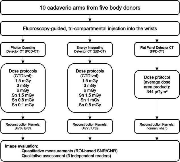
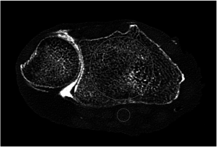
Methods: Following ethics committee approval, ten cadaveric wrists were injected with diluted iodinated contrast agent. CT arthrographies using PCD-, EID-, and FPD-CT were performed. Six dose protocols between 0.1 mGy (using a tin filter) and 6 mGy, ultrahigh-resolution-mode, and two reconstruction kernels were used for the PCD-CT and EID-CT. FPD-CT images were reconstructed using a "normal" and "sharp" kernel. Signal-to-noise ratios (SNR) and contrast-to-noise ratios (CNR) were calculated and analyzed using analysis of variance (ANOVA) and post hoc tests. Three blinded radiologists independently rated image quality concerning trabecular, cartilage, and intrinsic structures. Intraclass correlation coefficients (ICC) were calculated, followed by a Friedman and post hoc test.
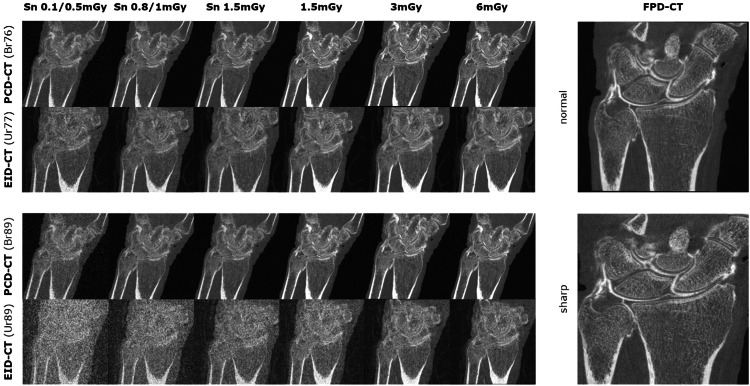
Results: At 1.5 mGy, 3 mGy, and 6 mGy with the Br89 kernel, the PCD-CT yielded up to 2.35 times higher SNR and up to 7 times higher CNR than dose-equivalent and higher dose EID-CT scans. Subjective ratings favored the PCD-CT over the EID-CT and occasionally the FPD-CT, with a combined ICC of 0.942. Applying sharper kernels, SNR did not differ significantly between the PCD-CT (1.5 mGy, 3 mGy, and 6 mGy) and the FPD-CT.
Conclusion: Using sharp kernels, the PCD-CT provided superior image quality to the EID-CT and achieved comparable or better quality than the FPD at certain parameters. Thus, the PCD-CT could be considered a possible alternative in clinical routine for evaluating wrist injuries.
Relevance statement: This study demonstrates the potential of the PCD-CT as a valuable tool in diagnosing wrist injuries. Its superior image quality compared to the EID-CT can increase confidence in diagnosing subtle bone pathologies and additionally yields the possibility of radiation exposure reduction.
Key points: The technical advantages of the PCD-CT allow for dose reduction while generating high-quality images. PCD-CT showed superior image quality over EID-CT and was comparable to the FPD-CT. PDC-CT offers improved visualization of fine joint structures in wrist arthrography and should be considered in clinical routine.

 求助内容:
求助内容: 应助结果提醒方式:


