Xin Bai, Lin Lu, Anli Tong, Jianhua Deng, Lili Xu, Xiaoxiao Zhang, Jiahui Zhang, Li Chen, Qianyu Peng, Erjia Guo, Yongfei Wu, Yun Wang, Kai Xu, Chao Zhang, Xi Zhao, Zhengyu Jin, Gumuyang Zhang, Hao Sun
{"title":"Virtual noncontrast images of adrenal lesions: a photon-counting CT prospective study.","authors":"Xin Bai, Lin Lu, Anli Tong, Jianhua Deng, Lili Xu, Xiaoxiao Zhang, Jiahui Zhang, Li Chen, Qianyu Peng, Erjia Guo, Yongfei Wu, Yun Wang, Kai Xu, Chao Zhang, Xi Zhao, Zhengyu Jin, Gumuyang Zhang, Hao Sun","doi":"10.1186/s41747-025-00621-x","DOIUrl":null,"url":null,"abstract":"<p><strong>Background: </strong>The value of virtual noncontrast (VNC) images from photon-counting computed tomography (PCCT) for evaluating adrenal lesions and diagnosing adrenal adenomas remains to be clarified.</p><p><strong>Materials and methods: </strong>Participants with adrenal masses who underwent unenhanced and portal venous phase PCCT were prospectively included. Portal-venous phase images were reconstructed using conventional VNC (VNC<sub>Conv</sub>) and PureCalcium VNC (VNC<sub>PC</sub>). We measured two-dimensional (2D) attenuation of adrenal masses at their largest slice on true noncontrast (TNC), VNC<sub>Conv</sub>, and VNC<sub>PC</sub> images. Three-dimensional (3D) attenuation and radiomic features of adrenal masses were semiautomatically extracted. These parameters were statistically compared, and diagnostic performance for adenomas was evaluated.</p><p><strong>Results: </strong>The study included 54 participants (27 females, mean age 45.3 years) with 68 adrenal lesions. Attenuation values on VNC were higher than those on TNC. TNC, VNC<sub>Conv</sub>, and VNC<sub>PC</sub> attenuation values did not differ between 2D and 3D measurements. The intraclass correlation coefficients of first-order, shape, and texture features between TNC and VNC were 0.671, 0.822, and 0.616, respectively. The sensitivity and specificity of the proposed thresholds (VNC<sub>Conv</sub> 25 HU, VNC<sub>PC</sub> 20 HU) were higher than those of the previously established threshold of 10 HU in diagnosing adenomas. There was no significant difference between VNC<sub>Conv</sub> and VNC<sub>PC</sub> in diagnosing adenomas (area under the receiver operating characteristic curve: 0.841 versus 0.838, p = 0.873).</p><p><strong>Conclusion: </strong>VNC algorithms from PCCT overestimated CT attenuation of adrenal lesions. Higher thresholds showed better diagnostic performance for discriminating adrenal adenomas from non-adenomas than the established 10 HU.</p><p><strong>Relevance statement: </strong>We investigated the application of VNC images from PCCT in adrenal disease. On VNC images, higher thresholds, superior to the accepted 10 HU, are needed for discriminating adenomas from non-adenomas, reducing the need for secondary examinations.</p><p><strong>Key points: </strong>This study investigated the value of VNC images from PCCT in adrenal lesions. VNC reconstruction overestimated the CT attenuation of adrenal lesions. Higher thresholds on VNC images were superior to the accepted 10 HU for differentiating adenomas from non-adenomas.</p>","PeriodicalId":36926,"journal":{"name":"European Radiology Experimental","volume":"9 1","pages":"82"},"PeriodicalIF":3.6000,"publicationDate":"2025-08-29","publicationTypes":"Journal Article","fieldsOfStudy":null,"isOpenAccess":false,"openAccessPdf":"https://www.ncbi.nlm.nih.gov/pmc/articles/PMC12396999/pdf/","citationCount":"0","resultStr":null,"platform":"Semanticscholar","paperid":null,"PeriodicalName":"European Radiology Experimental","FirstCategoryId":"1085","ListUrlMain":"https://doi.org/10.1186/s41747-025-00621-x","RegionNum":0,"RegionCategory":null,"ArticlePicture":[],"TitleCN":null,"AbstractTextCN":null,"PMCID":null,"EPubDate":"","PubModel":"","JCR":"Q1","JCRName":"RADIOLOGY, NUCLEAR MEDICINE & MEDICAL IMAGING","Score":null,"Total":0}
引用次数: 0
Abstract
Background: The value of virtual noncontrast (VNC) images from photon-counting computed tomography (PCCT) for evaluating adrenal lesions and diagnosing adrenal adenomas remains to be clarified.
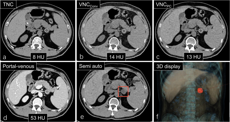
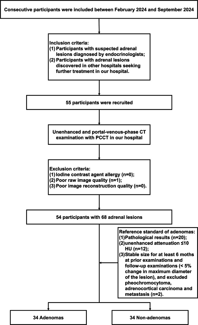
Materials and methods: Participants with adrenal masses who underwent unenhanced and portal venous phase PCCT were prospectively included. Portal-venous phase images were reconstructed using conventional VNC (VNCConv) and PureCalcium VNC (VNCPC). We measured two-dimensional (2D) attenuation of adrenal masses at their largest slice on true noncontrast (TNC), VNCConv, and VNCPC images. Three-dimensional (3D) attenuation and radiomic features of adrenal masses were semiautomatically extracted. These parameters were statistically compared, and diagnostic performance for adenomas was evaluated.
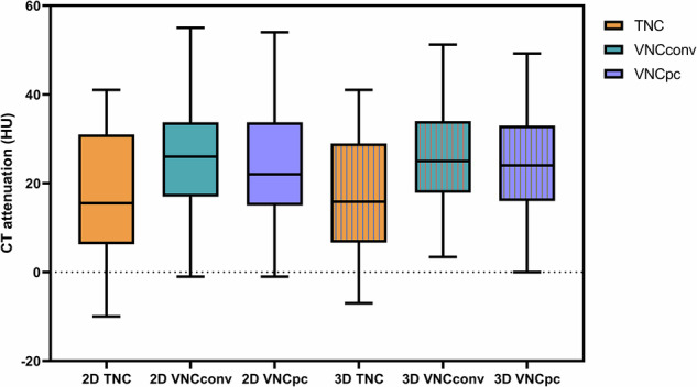
Results: The study included 54 participants (27 females, mean age 45.3 years) with 68 adrenal lesions. Attenuation values on VNC were higher than those on TNC. TNC, VNCConv, and VNCPC attenuation values did not differ between 2D and 3D measurements. The intraclass correlation coefficients of first-order, shape, and texture features between TNC and VNC were 0.671, 0.822, and 0.616, respectively. The sensitivity and specificity of the proposed thresholds (VNCConv 25 HU, VNCPC 20 HU) were higher than those of the previously established threshold of 10 HU in diagnosing adenomas. There was no significant difference between VNCConv and VNCPC in diagnosing adenomas (area under the receiver operating characteristic curve: 0.841 versus 0.838, p = 0.873).
Conclusion: VNC algorithms from PCCT overestimated CT attenuation of adrenal lesions. Higher thresholds showed better diagnostic performance for discriminating adrenal adenomas from non-adenomas than the established 10 HU.
Relevance statement: We investigated the application of VNC images from PCCT in adrenal disease. On VNC images, higher thresholds, superior to the accepted 10 HU, are needed for discriminating adenomas from non-adenomas, reducing the need for secondary examinations.
Key points: This study investigated the value of VNC images from PCCT in adrenal lesions. VNC reconstruction overestimated the CT attenuation of adrenal lesions. Higher thresholds on VNC images were superior to the accepted 10 HU for differentiating adenomas from non-adenomas.

 求助内容:
求助内容: 应助结果提醒方式:


