{"title":"Atypical presentation of a large posterior falx meningioma involving the parafalcine region in a 78-year-female: A case report.","authors":"Nadeem AlSabea, Samar Syeda, Marina Gubran, Viktoriya Gibatova, Rahul Sharma, Anjiya Aswani","doi":"10.12998/wjcc.v13.i25.108429","DOIUrl":null,"url":null,"abstract":"<p><strong>Background: </strong>Meningiomas represent the most common primary intracranial tumor in adults. The majority of meningiomas are indolent, benign, and sporadic in nature. The incidence of meningiomas is directly proportional to the age, peaking around 65 years. The presenting symptomatology of intracranial meningiomas is mainly dependent on their anatomical location, as with the majority of brain tumors. Surgical resection and radiation therapy remain the treatment modality for meningiomas of all grades.</p><p><strong>Case summary: </strong>We present a case describing a 78-year-old female who came in following a ground level fall. The primary assessment was notable for a history of similar recurrent falls and subtle left-sided peripheral visual field loss. Further neurological examination was otherwise largely unremarkable. A computed tomography scan of the head revealed a large extra-axial mass located along the posterior aspect of the falx. Follow-up magnetic resonance imaging confirmed a lesion measuring around 6.6 cm × 4.2 cm × 5.5 cm. A partial surgical resection of the right-sided portion of the lesion was performed. Complete resection was limited by insufficient visualization and challenges with hemostatic control of the left parafalcine region. Further histopathological analysis confirmed a fibrous meningioma with focal necrosis, consistent with World Health Organization Grade 2 classification. She was subsequently scheduled for outpatient follow-up to assess the residual tumor management.</p><p><strong>Conclusion: </strong>This case highlights the importance of maintaining a high index of suspicion for intracranial pathology in elderly patients with nonspecific presentation.</p>","PeriodicalId":23912,"journal":{"name":"World Journal of Clinical Cases","volume":"13 25","pages":"108429"},"PeriodicalIF":1.0000,"publicationDate":"2025-09-06","publicationTypes":"Journal Article","fieldsOfStudy":null,"isOpenAccess":false,"openAccessPdf":"https://www.ncbi.nlm.nih.gov/pmc/articles/PMC12243921/pdf/","citationCount":"0","resultStr":null,"platform":"Semanticscholar","paperid":null,"PeriodicalName":"World Journal of Clinical Cases","FirstCategoryId":"3","ListUrlMain":"https://doi.org/10.12998/wjcc.v13.i25.108429","RegionNum":4,"RegionCategory":"医学","ArticlePicture":[],"TitleCN":null,"AbstractTextCN":null,"PMCID":null,"EPubDate":"","PubModel":"","JCR":"Q3","JCRName":"MEDICINE, GENERAL & INTERNAL","Score":null,"Total":0}
引用次数: 0
Abstract
Background: Meningiomas represent the most common primary intracranial tumor in adults. The majority of meningiomas are indolent, benign, and sporadic in nature. The incidence of meningiomas is directly proportional to the age, peaking around 65 years. The presenting symptomatology of intracranial meningiomas is mainly dependent on their anatomical location, as with the majority of brain tumors. Surgical resection and radiation therapy remain the treatment modality for meningiomas of all grades.
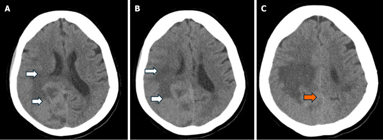
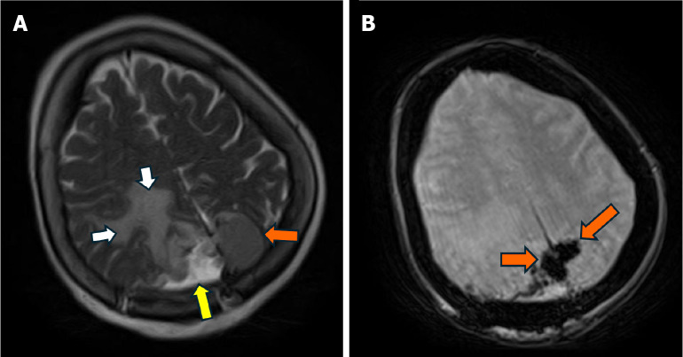
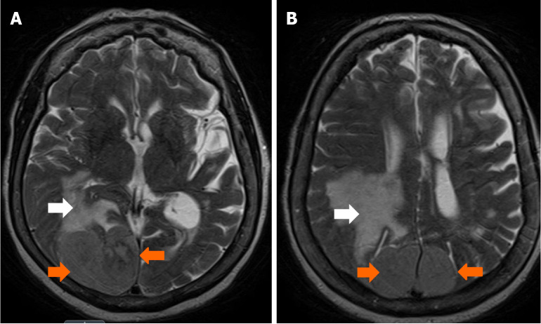
Case summary: We present a case describing a 78-year-old female who came in following a ground level fall. The primary assessment was notable for a history of similar recurrent falls and subtle left-sided peripheral visual field loss. Further neurological examination was otherwise largely unremarkable. A computed tomography scan of the head revealed a large extra-axial mass located along the posterior aspect of the falx. Follow-up magnetic resonance imaging confirmed a lesion measuring around 6.6 cm × 4.2 cm × 5.5 cm. A partial surgical resection of the right-sided portion of the lesion was performed. Complete resection was limited by insufficient visualization and challenges with hemostatic control of the left parafalcine region. Further histopathological analysis confirmed a fibrous meningioma with focal necrosis, consistent with World Health Organization Grade 2 classification. She was subsequently scheduled for outpatient follow-up to assess the residual tumor management.
Conclusion: This case highlights the importance of maintaining a high index of suspicion for intracranial pathology in elderly patients with nonspecific presentation.
背景:脑膜瘤是成人最常见的原发性颅内肿瘤。大多数脑膜瘤是惰性的,良性的,散发性的。脑膜瘤的发病率与年龄成正比,在65岁左右达到高峰。与大多数脑肿瘤一样,颅内脑膜瘤的表现主要取决于其解剖位置。手术切除和放射治疗仍然是所有级别脑膜瘤的治疗方式。病例总结:我们报告了一个78岁的女性病例,她在地面坠落后入院。最初的评估是值得注意的历史类似的复发性跌倒和轻微的左侧周边视野丧失。除此之外,进一步的神经学检查基本上没有什么特别之处。头部的计算机断层扫描显示一个大的轴外肿块位于镰的后侧。后续磁共振成像证实病变大小约为6.6 cm × 4.2 cm × 5.5 cm。手术切除病灶右侧部分。完全切除是有限的,由于不充分的可视化和挑战止血控制左镰旁区。进一步的组织病理学分析证实为纤维性脑膜瘤伴局灶性坏死,符合世界卫生组织分级2级。随后安排门诊随访以评估残余肿瘤的处理情况。结论:本病例强调了对非特异性表现的老年患者颅内病理保持高怀疑指数的重要性。
期刊介绍:
The World Journal of Clinical Cases (WJCC) is a high-quality, peer reviewed, open-access journal. The primary task of WJCC is to rapidly publish high-quality original articles, reviews, editorials, and case reports in the field of clinical cases. In order to promote productive academic communication, the peer review process for the WJCC is transparent; to this end, all published manuscripts are accompanied by the anonymized reviewers’ comments as well as the authors’ responses. The primary aims of the WJCC are to improve diagnostic, therapeutic and preventive modalities and the skills of clinicians and to guide clinical practice in clinical cases.

 求助内容:
求助内容: 应助结果提醒方式:


