Mesenchymal stem cell-derived lncRNAs NKILA contributes to stemness and chemoresistance by fatty acid oxidation in gastric cancer via miR-485-5p/STAT3.
{"title":"Mesenchymal stem cell-derived lncRNAs NKILA contributes to stemness and chemoresistance by fatty acid oxidation in gastric cancer <i>via</i> miR-485-5p/STAT3.","authors":"Xiao-Juan Lyu, Lin Zhou, Xu-Mian Jiang, Dan Zheng","doi":"10.4251/wjgo.v17.i8.105006","DOIUrl":null,"url":null,"abstract":"<p><strong>Background: </strong>Gastric cancer (GC) is a type of cancer which causes high cancer-related mortality. Surgical operation and systematic chemical therapies are primary choices for the treatment of GC patients with advanced stages, however, the 5-year overall survival is only around 30%.</p><p><strong>Aim: </strong>To investigate the role of mesenchymal stem cell (MSC)-derived long non-coding RNAs (lncRNA) NKILA in fatty acid oxidation and chemoresistance in GC cells, mediated through the miR-485-5p/STAT3 pathway.</p><p><strong>Methods: </strong>GC cell lines (AGS and MKN45) were co-cultured with human bone marrow-derived MSCs were cultured. The MSC identity was confirmed by flow cytometry (CD73, CD90, CD105 > 95% positive, CD34, CD45 negative). Co-culture of GC cells and MSCs was performed in Transwell plates, where MSCs were placed in the upper chamber and GC cells in the lower chamber for 72 hours. For transfections, pcDNA-NKILA vectors, shSTAT3, and miR-485-5p mimics were utilized. Colony formation, apoptosis assays (Annexin V/PI staining), sphere formation, and flow cytometry were performed to evaluate cell proliferation, stemness, and chemoresistance. qPCR was used to analyze gene expression (Sox2, Oct4, CD133, LIN28, NKILA), and Western blotting assessed protein levels of stemness markers. Luciferase reporter assays were conducted to confirm miR-485-5p/STAT3 interactions, and biotin-labeled RNA pulldown was used to assess RNA-protein binding. Fatty acid oxidation was evaluated using a CPT1 activity assay and β-oxidation rate detection. ATP levels were measured to assess the energetic status of GC cells. Clinical GC tissue samples were collected from patients at our hospital for validation.</p><p><strong>Results: </strong>MSCs were found to enhance the stemness and chemoresistance of GC cells. Co-culturing MKN45 and AGS cells with MSCs significantly increased sphere-forming ability and the expression of key cancer stem cell markers (SOX2, Oct4, LIN28, CD133), indicating that MSCs promote stem-like properties. Flow cytometry confirmed an enrichment of CD44+ and CD133+ subpopulations in MSC-treated GC cells. Additionally, MSC co-culture reduced chemotherapy-induced apoptosis and enhanced cell proliferation, suggesting a protective role in chemotherapy resistance. MSC-derived lncRNA NKILA further promoted stemness and chemoresistance, enhancing expression of stem cell markers and protecting cells from oxaliplatin and 5-FU-induced apoptosis. MSC co-culture also induced fatty acid oxidation in GC cells, as shown by increased CPT1 activity, β-oxidation rates, and ATP levels. NKILA mediated these effects by upregulating STAT3, which was confirmed to regulate fatty acid oxidation and chemoresistance. NKILA's interaction with miR-485-5p further promoted STAT3 expression and fatty acid oxidation, reinforcing its role in maintaining stemness and enhancing chemoresistance.</p><p><strong>Conclusion: </strong>MSCs enhance the stemness and chemoresistance of GC cells by secreting lncRNA NKILA, which promotes fatty acid oxidation through STAT3 activation. NKILA modulates the miR-485-5p/STAT3 axis, thereby increasing energy metabolism and supporting cancer stem cell properties. Targeting NKILA or the miR-485-5p/STAT3 pathway offers potential therapeutic strategies to overcome chemoresistance in GC.</p>","PeriodicalId":23762,"journal":{"name":"World Journal of Gastrointestinal Oncology","volume":"17 8","pages":"105006"},"PeriodicalIF":2.5000,"publicationDate":"2025-08-15","publicationTypes":"Journal Article","fieldsOfStudy":null,"isOpenAccess":false,"openAccessPdf":"https://www.ncbi.nlm.nih.gov/pmc/articles/PMC12362598/pdf/","citationCount":"0","resultStr":null,"platform":"Semanticscholar","paperid":null,"PeriodicalName":"World Journal of Gastrointestinal Oncology","FirstCategoryId":"3","ListUrlMain":"https://doi.org/10.4251/wjgo.v17.i8.105006","RegionNum":4,"RegionCategory":"医学","ArticlePicture":[],"TitleCN":null,"AbstractTextCN":null,"PMCID":null,"EPubDate":"","PubModel":"","JCR":"Q2","JCRName":"GASTROENTEROLOGY & HEPATOLOGY","Score":null,"Total":0}
引用次数: 0
Abstract
Background: Gastric cancer (GC) is a type of cancer which causes high cancer-related mortality. Surgical operation and systematic chemical therapies are primary choices for the treatment of GC patients with advanced stages, however, the 5-year overall survival is only around 30%.
Aim: To investigate the role of mesenchymal stem cell (MSC)-derived long non-coding RNAs (lncRNA) NKILA in fatty acid oxidation and chemoresistance in GC cells, mediated through the miR-485-5p/STAT3 pathway.
Methods: GC cell lines (AGS and MKN45) were co-cultured with human bone marrow-derived MSCs were cultured. The MSC identity was confirmed by flow cytometry (CD73, CD90, CD105 > 95% positive, CD34, CD45 negative). Co-culture of GC cells and MSCs was performed in Transwell plates, where MSCs were placed in the upper chamber and GC cells in the lower chamber for 72 hours. For transfections, pcDNA-NKILA vectors, shSTAT3, and miR-485-5p mimics were utilized. Colony formation, apoptosis assays (Annexin V/PI staining), sphere formation, and flow cytometry were performed to evaluate cell proliferation, stemness, and chemoresistance. qPCR was used to analyze gene expression (Sox2, Oct4, CD133, LIN28, NKILA), and Western blotting assessed protein levels of stemness markers. Luciferase reporter assays were conducted to confirm miR-485-5p/STAT3 interactions, and biotin-labeled RNA pulldown was used to assess RNA-protein binding. Fatty acid oxidation was evaluated using a CPT1 activity assay and β-oxidation rate detection. ATP levels were measured to assess the energetic status of GC cells. Clinical GC tissue samples were collected from patients at our hospital for validation.
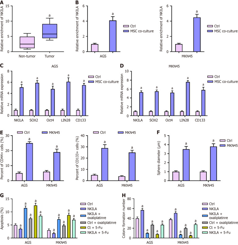
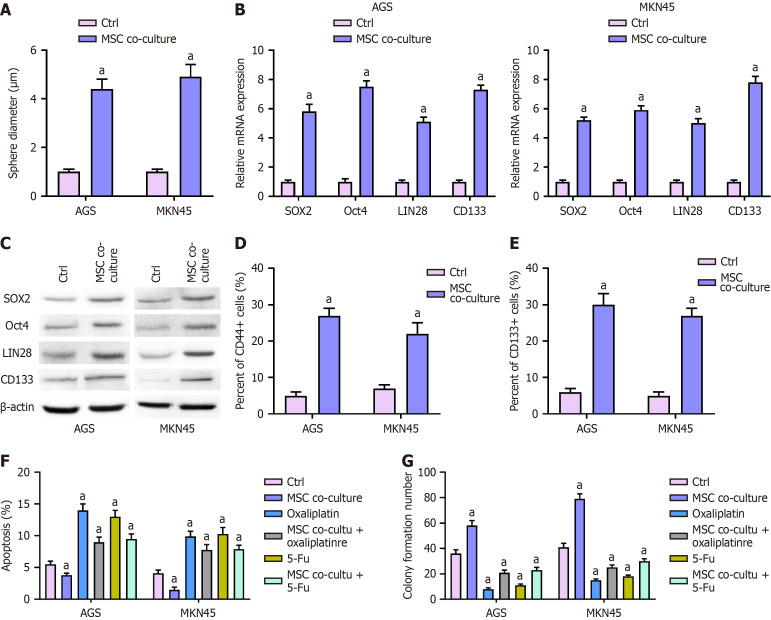
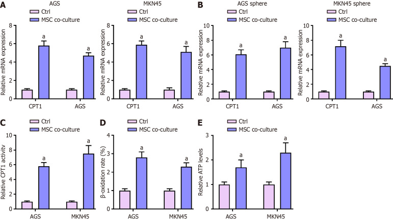
Results: MSCs were found to enhance the stemness and chemoresistance of GC cells. Co-culturing MKN45 and AGS cells with MSCs significantly increased sphere-forming ability and the expression of key cancer stem cell markers (SOX2, Oct4, LIN28, CD133), indicating that MSCs promote stem-like properties. Flow cytometry confirmed an enrichment of CD44+ and CD133+ subpopulations in MSC-treated GC cells. Additionally, MSC co-culture reduced chemotherapy-induced apoptosis and enhanced cell proliferation, suggesting a protective role in chemotherapy resistance. MSC-derived lncRNA NKILA further promoted stemness and chemoresistance, enhancing expression of stem cell markers and protecting cells from oxaliplatin and 5-FU-induced apoptosis. MSC co-culture also induced fatty acid oxidation in GC cells, as shown by increased CPT1 activity, β-oxidation rates, and ATP levels. NKILA mediated these effects by upregulating STAT3, which was confirmed to regulate fatty acid oxidation and chemoresistance. NKILA's interaction with miR-485-5p further promoted STAT3 expression and fatty acid oxidation, reinforcing its role in maintaining stemness and enhancing chemoresistance.
Conclusion: MSCs enhance the stemness and chemoresistance of GC cells by secreting lncRNA NKILA, which promotes fatty acid oxidation through STAT3 activation. NKILA modulates the miR-485-5p/STAT3 axis, thereby increasing energy metabolism and supporting cancer stem cell properties. Targeting NKILA or the miR-485-5p/STAT3 pathway offers potential therapeutic strategies to overcome chemoresistance in GC.
期刊介绍:
The World Journal of Gastrointestinal Oncology (WJGO) is a leading academic journal devoted to reporting the latest, cutting-edge research progress and findings of basic research and clinical practice in the field of gastrointestinal oncology.

 求助内容:
求助内容: 应助结果提醒方式:


