{"title":"Correlation between baseline magnetic resonance imaging features and serum carcinoembryonic antigen levels in patients with primary rectal cancer.","authors":"Peng Wang, Wen-Na Zhao, Jun Han, Kai-Xuan Wang, Xiao-Feng Yang, Yi-Juan Huang","doi":"10.4251/wjgo.v17.i8.108016","DOIUrl":null,"url":null,"abstract":"<p><strong>Background: </strong>Serum carcinoembryonic antigen (CEA) levels and magnetic resonance imaging (MRI) findings are widely used for the diagnosis and treatment of rectal cancer; however, research investigating their correlation remains limited.</p><p><strong>Aim: </strong>To investigate the correlation between baseline MRI features and serum CEA levels in patients diagnosed with primary rectal cancer.</p><p><strong>Methods: </strong>Eighty patients (age: 42-78 years) diagnosed with primary rectal cancer were enrolled. Baseline MRI examinations were performed to evaluate tumor size, T stage, circumferential resection margin status, extramural vascular invasion (EMVI), and lymph node metastasis. Serum CEA levels were concurrently measured. Statistical methods were used to analyze correlations.</p><p><strong>Results: </strong>Tumor size, T stage, EMVI, and lymph node metastasis were significantly correlated with serum CEA levels (<i>P</i> < 0.05). Multivariate analysis identified T stage and lymph node metastasis as independent factors influencing serum CEA levels.</p><p><strong>Conclusion: </strong>This study confirmed the correlation between baseline MRI features and serum CEA levels in patients with primary rectal cancer, highlighting their potential utility for precise diagnosis, staging, and prognostic evaluation.</p>","PeriodicalId":23762,"journal":{"name":"World Journal of Gastrointestinal Oncology","volume":"17 8","pages":"108016"},"PeriodicalIF":2.5000,"publicationDate":"2025-08-15","publicationTypes":"Journal Article","fieldsOfStudy":null,"isOpenAccess":false,"openAccessPdf":"https://www.ncbi.nlm.nih.gov/pmc/articles/PMC12362563/pdf/","citationCount":"0","resultStr":null,"platform":"Semanticscholar","paperid":null,"PeriodicalName":"World Journal of Gastrointestinal Oncology","FirstCategoryId":"3","ListUrlMain":"https://doi.org/10.4251/wjgo.v17.i8.108016","RegionNum":4,"RegionCategory":"医学","ArticlePicture":[],"TitleCN":null,"AbstractTextCN":null,"PMCID":null,"EPubDate":"","PubModel":"","JCR":"Q2","JCRName":"GASTROENTEROLOGY & HEPATOLOGY","Score":null,"Total":0}
引用次数: 0
Abstract
Background: Serum carcinoembryonic antigen (CEA) levels and magnetic resonance imaging (MRI) findings are widely used for the diagnosis and treatment of rectal cancer; however, research investigating their correlation remains limited.
Aim: To investigate the correlation between baseline MRI features and serum CEA levels in patients diagnosed with primary rectal cancer.
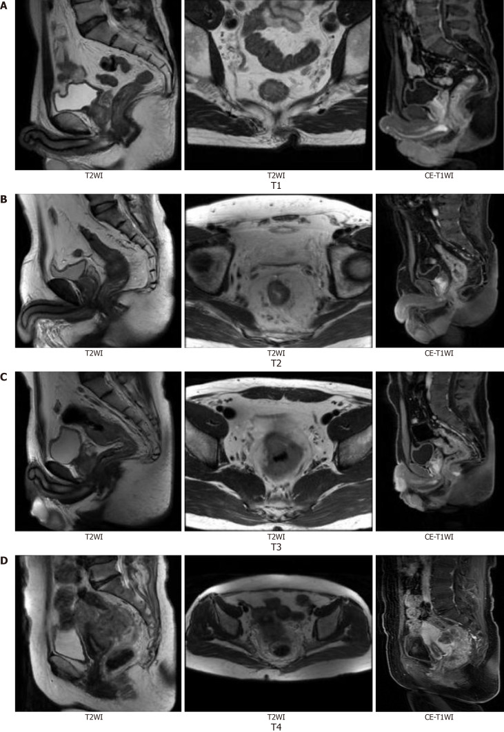
Methods: Eighty patients (age: 42-78 years) diagnosed with primary rectal cancer were enrolled. Baseline MRI examinations were performed to evaluate tumor size, T stage, circumferential resection margin status, extramural vascular invasion (EMVI), and lymph node metastasis. Serum CEA levels were concurrently measured. Statistical methods were used to analyze correlations.
Results: Tumor size, T stage, EMVI, and lymph node metastasis were significantly correlated with serum CEA levels (P < 0.05). Multivariate analysis identified T stage and lymph node metastasis as independent factors influencing serum CEA levels.
Conclusion: This study confirmed the correlation between baseline MRI features and serum CEA levels in patients with primary rectal cancer, highlighting their potential utility for precise diagnosis, staging, and prognostic evaluation.
期刊介绍:
The World Journal of Gastrointestinal Oncology (WJGO) is a leading academic journal devoted to reporting the latest, cutting-edge research progress and findings of basic research and clinical practice in the field of gastrointestinal oncology.

 求助内容:
求助内容: 应助结果提醒方式:


