Peng Wang, Jun Han, Wen-Na Zhao, Fan Wu, Sheng-He Zhang, Yi-Juan Huang
{"title":"Tumor markers and multimodal magnetic resonance imaging in predicting rectal cancer stage and differentiation.","authors":"Peng Wang, Jun Han, Wen-Na Zhao, Fan Wu, Sheng-He Zhang, Yi-Juan Huang","doi":"10.4251/wjgo.v17.i8.108007","DOIUrl":null,"url":null,"abstract":"<p><strong>Background: </strong>Rectal cancer is one of the common digestive system malignant tumors around the world. Its early diagnosis and staging are crucial for rectal cancer treatment and prognosis. In recent years, tumor markers have gradually received attention in early screening, treatment monitoring and prognostic evaluation of cancer, but their predictive role in rectal cancer staging and differentiation is still unclear.</p><p><strong>Aim: </strong>To assess the prognostic value of tumor markers alpha-fetoprotein (AFP) cancer antigen 72-4 (CA72-4), carbohydrate antigen 19-9 (CA19-9), and carcinoembryonic antigen (CEA), alongside multimodal magnetic resonance imaging (MRI), for staging and differentiating rectal cancer in patients.</p><p><strong>Methods: </strong>This study retrospectively analyzed 167 patients with rectal cancer who were treated at our institution from January 2020 to December 2024. Each patient underwent serological testing and multimodal MRI for diagnosis. Histopathological examination after surgical resection or imaging based on follow-up was used as the gold standard. According to the T stage and differentiation degree, patients were divided into low stage group (T1-T2) and high stage group (T3-T4). In addition, they were divided into low-differentiation groups and high-differentiation groups according to their differentiation degree. We compared the accuracy, sensitivity and specificity of tumor marker levels and MRI in rectal cancer stage and differentiation.</p><p><strong>Results: </strong>The study's findings indicate that in the context of rectal cancer T staging, there is substantial concordance between MRI and clinicopathological assessments, with a Kappa coefficient of 0.789 (<i>P</i> < 0.001). Similarly, for various degrees of tumor differentiation, MRI and clinicopathological evaluations demonstrated substantial agreement, with a Kappa coefficient of 0.651 (<i>P</i> < 0.001). Notably, the concentrations of tumor markers CA19-9, CA72-4, CEA, and AFP were significantly elevated in the T3-T4 stage compared to the T1-T2 stage. Furthermore, these markers were significantly higher in the low-differentiation group compared to the high-differentiation group (<i>P</i> < 0.05). The combined use of tumor markers and MRI for preoperative T staging of rectal cancer yielded a diagnostic sensitivity of 93.7% and a specificity of 94.6%, as evidenced by the receiver operating characteristic analysis, with an area under the curve of 0.947. For tumor differentiation, the diagnostic sensitivity and specificity were 93.6% and 97.1%, respectively, with an area under the curve of 0.978 (95% confidence interval: 0.946-1.000), surpassing the accuracy of individual detection methods.</p><p><strong>Conclusion: </strong>The CA19-9, CA72-4, CEA and AFP tumor markers combined with multimodal MRI have high sensitivity and specificity in diagnosing rectal cancer stage and differentiation. Their diagnostic efficacy is significantly better than that of single tests, which can effectively improve the predictive ability of rectal cancer stage and differentiation, provide a more reliable diagnostic reference for clinical practice, and have important clinical significance.</p>","PeriodicalId":23762,"journal":{"name":"World Journal of Gastrointestinal Oncology","volume":"17 8","pages":"108007"},"PeriodicalIF":2.5000,"publicationDate":"2025-08-15","publicationTypes":"Journal Article","fieldsOfStudy":null,"isOpenAccess":false,"openAccessPdf":"https://www.ncbi.nlm.nih.gov/pmc/articles/PMC12362569/pdf/","citationCount":"0","resultStr":null,"platform":"Semanticscholar","paperid":null,"PeriodicalName":"World Journal of Gastrointestinal Oncology","FirstCategoryId":"3","ListUrlMain":"https://doi.org/10.4251/wjgo.v17.i8.108007","RegionNum":4,"RegionCategory":"医学","ArticlePicture":[],"TitleCN":null,"AbstractTextCN":null,"PMCID":null,"EPubDate":"","PubModel":"","JCR":"Q2","JCRName":"GASTROENTEROLOGY & HEPATOLOGY","Score":null,"Total":0}
引用次数: 0
Abstract
Background: Rectal cancer is one of the common digestive system malignant tumors around the world. Its early diagnosis and staging are crucial for rectal cancer treatment and prognosis. In recent years, tumor markers have gradually received attention in early screening, treatment monitoring and prognostic evaluation of cancer, but their predictive role in rectal cancer staging and differentiation is still unclear.
Aim: To assess the prognostic value of tumor markers alpha-fetoprotein (AFP) cancer antigen 72-4 (CA72-4), carbohydrate antigen 19-9 (CA19-9), and carcinoembryonic antigen (CEA), alongside multimodal magnetic resonance imaging (MRI), for staging and differentiating rectal cancer in patients.
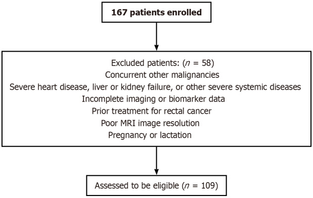
Methods: This study retrospectively analyzed 167 patients with rectal cancer who were treated at our institution from January 2020 to December 2024. Each patient underwent serological testing and multimodal MRI for diagnosis. Histopathological examination after surgical resection or imaging based on follow-up was used as the gold standard. According to the T stage and differentiation degree, patients were divided into low stage group (T1-T2) and high stage group (T3-T4). In addition, they were divided into low-differentiation groups and high-differentiation groups according to their differentiation degree. We compared the accuracy, sensitivity and specificity of tumor marker levels and MRI in rectal cancer stage and differentiation.
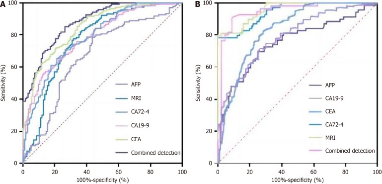
Results: The study's findings indicate that in the context of rectal cancer T staging, there is substantial concordance between MRI and clinicopathological assessments, with a Kappa coefficient of 0.789 (P < 0.001). Similarly, for various degrees of tumor differentiation, MRI and clinicopathological evaluations demonstrated substantial agreement, with a Kappa coefficient of 0.651 (P < 0.001). Notably, the concentrations of tumor markers CA19-9, CA72-4, CEA, and AFP were significantly elevated in the T3-T4 stage compared to the T1-T2 stage. Furthermore, these markers were significantly higher in the low-differentiation group compared to the high-differentiation group (P < 0.05). The combined use of tumor markers and MRI for preoperative T staging of rectal cancer yielded a diagnostic sensitivity of 93.7% and a specificity of 94.6%, as evidenced by the receiver operating characteristic analysis, with an area under the curve of 0.947. For tumor differentiation, the diagnostic sensitivity and specificity were 93.6% and 97.1%, respectively, with an area under the curve of 0.978 (95% confidence interval: 0.946-1.000), surpassing the accuracy of individual detection methods.
Conclusion: The CA19-9, CA72-4, CEA and AFP tumor markers combined with multimodal MRI have high sensitivity and specificity in diagnosing rectal cancer stage and differentiation. Their diagnostic efficacy is significantly better than that of single tests, which can effectively improve the predictive ability of rectal cancer stage and differentiation, provide a more reliable diagnostic reference for clinical practice, and have important clinical significance.
期刊介绍:
The World Journal of Gastrointestinal Oncology (WJGO) is a leading academic journal devoted to reporting the latest, cutting-edge research progress and findings of basic research and clinical practice in the field of gastrointestinal oncology.
 求助内容:
求助内容: 应助结果提醒方式:


