{"title":"Altered brain structure age gap estimation in major depressive disorder patients with and without anhedonia: a machine learning-based study.","authors":"Qingli Mu, Kejing Zhang, Yue Chen, Yuwei Xu, Shaohua Hu, Manli Huang, Peng Zhang, Dong Cui, Shaojia Lu","doi":"10.1038/s41398-025-03555-5","DOIUrl":null,"url":null,"abstract":"<p><p>Previous studies have found that major depressive disorder (MDD) may accelerate overall structural brain aging. Nevertheless, it still remains unknown whether anhedonia, a critical negative prognostic indicator in MDD, further leads to advanced brain aging in specific regions. A total of 31 MDD with anhedonia (MDD-WA), 41 MDD without anhedonia (MDD-WoA), and 43 healthy controls (HCs) were recruited in this study. The difference between brain structure age (BSA) applied by support vector regression (SVR) and chronological age was calculated to derive the brain structure age gap estimation (BSAGE). Analyses of covariance (ANCOVAs) and intergroup comparisons were performed to obtain brain regions with significant BSAGE differences among three groups. Moreover, a support vector machine (SVM) classification model was used to verify the diagnostic value of altered BSAGE. ANCOVAs revealed significant BSAGE differences among three groups in the bilateral putamen (PU), left cerebellar white matter (CB), left cuneus (CUN), left fusiform gyrus (FuG), left subcallosal area (SCA), left superior occipital gyrus (SOG), left triangular inferior frontal gyrus (IFG-Tri), right lateral ventricle (L-V), right superior frontal gyrus medial segment (SFG-SM), right opercular inferior frontal gyrus (IFG-Oper), right precuneus (pre-CUN), right posterior insula (INS-Post), and right superior temporal gyrus (STG). Compared to HCs, the MDD-WA group showed significant BSAGE increase in all of the aforementioned brain regions, while the MDD-WoA group showed limited BSAGE increase in the CB, FuG, and SCA of left hemisphere only. However, no significant difference was found between MDD-WA and MDD-WoA. The altered BSAGE values showed promising discriminatory performance with an area under the curve (AUC) of 0.944 in classifying MDD-WA and HCs. The current findings emphasize that MDD with anhedonia may exhibit more extensive advanced brain aging, primarily in the frontal-limbic system, temporal lobe, and parietal lobe.</p>","PeriodicalId":23278,"journal":{"name":"Translational Psychiatry","volume":"15 1","pages":"309"},"PeriodicalIF":6.2000,"publicationDate":"2025-08-22","publicationTypes":"Journal Article","fieldsOfStudy":null,"isOpenAccess":false,"openAccessPdf":"https://www.ncbi.nlm.nih.gov/pmc/articles/PMC12373830/pdf/","citationCount":"0","resultStr":null,"platform":"Semanticscholar","paperid":null,"PeriodicalName":"Translational Psychiatry","FirstCategoryId":"3","ListUrlMain":"https://doi.org/10.1038/s41398-025-03555-5","RegionNum":1,"RegionCategory":"医学","ArticlePicture":[],"TitleCN":null,"AbstractTextCN":null,"PMCID":null,"EPubDate":"","PubModel":"","JCR":"Q1","JCRName":"PSYCHIATRY","Score":null,"Total":0}
引用次数: 0
Abstract
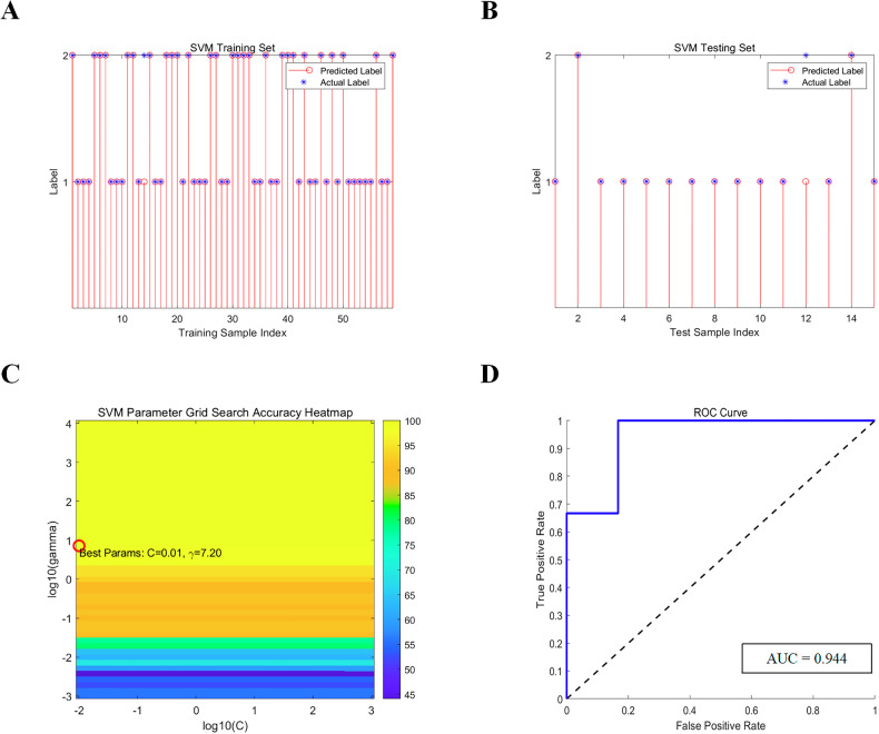
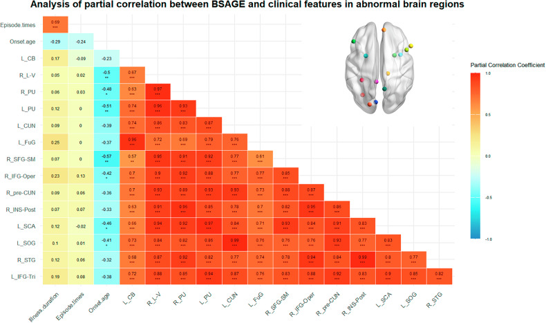
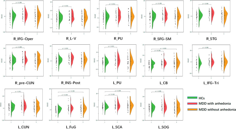
Previous studies have found that major depressive disorder (MDD) may accelerate overall structural brain aging. Nevertheless, it still remains unknown whether anhedonia, a critical negative prognostic indicator in MDD, further leads to advanced brain aging in specific regions. A total of 31 MDD with anhedonia (MDD-WA), 41 MDD without anhedonia (MDD-WoA), and 43 healthy controls (HCs) were recruited in this study. The difference between brain structure age (BSA) applied by support vector regression (SVR) and chronological age was calculated to derive the brain structure age gap estimation (BSAGE). Analyses of covariance (ANCOVAs) and intergroup comparisons were performed to obtain brain regions with significant BSAGE differences among three groups. Moreover, a support vector machine (SVM) classification model was used to verify the diagnostic value of altered BSAGE. ANCOVAs revealed significant BSAGE differences among three groups in the bilateral putamen (PU), left cerebellar white matter (CB), left cuneus (CUN), left fusiform gyrus (FuG), left subcallosal area (SCA), left superior occipital gyrus (SOG), left triangular inferior frontal gyrus (IFG-Tri), right lateral ventricle (L-V), right superior frontal gyrus medial segment (SFG-SM), right opercular inferior frontal gyrus (IFG-Oper), right precuneus (pre-CUN), right posterior insula (INS-Post), and right superior temporal gyrus (STG). Compared to HCs, the MDD-WA group showed significant BSAGE increase in all of the aforementioned brain regions, while the MDD-WoA group showed limited BSAGE increase in the CB, FuG, and SCA of left hemisphere only. However, no significant difference was found between MDD-WA and MDD-WoA. The altered BSAGE values showed promising discriminatory performance with an area under the curve (AUC) of 0.944 in classifying MDD-WA and HCs. The current findings emphasize that MDD with anhedonia may exhibit more extensive advanced brain aging, primarily in the frontal-limbic system, temporal lobe, and parietal lobe.
期刊介绍:
Psychiatry has suffered tremendously by the limited translational pipeline. Nobel laureate Julius Axelrod''s discovery in 1961 of monoamine reuptake by pre-synaptic neurons still forms the basis of contemporary antidepressant treatment. There is a grievous gap between the explosion of knowledge in neuroscience and conceptually novel treatments for our patients. Translational Psychiatry bridges this gap by fostering and highlighting the pathway from discovery to clinical applications, healthcare and global health. We view translation broadly as the full spectrum of work that marks the pathway from discovery to global health, inclusive. The steps of translation that are within the scope of Translational Psychiatry include (i) fundamental discovery, (ii) bench to bedside, (iii) bedside to clinical applications (clinical trials), (iv) translation to policy and health care guidelines, (v) assessment of health policy and usage, and (vi) global health. All areas of medical research, including — but not restricted to — molecular biology, genetics, pharmacology, imaging and epidemiology are welcome as they contribute to enhance the field of translational psychiatry.
 求助内容:
求助内容: 应助结果提醒方式:


