{"title":"Gastrointestinal schwannomas: A case series of 9 patients and literature review.","authors":"Server Sezgin Uludağ, Ergin Erginöz, Nazım Güreş, Zeynep Özdemir, Nuray Kepil, Şebnem Batur","doi":"10.47717/turkjsurg.2025.2025-5-28","DOIUrl":null,"url":null,"abstract":"<p><p>Gastrointestinal schwannomas are benign, slow-growing, rare tumors comprising 2-6% of all mesenchymal tumors of the gastrointestinal tract and 0.2% of all gastric neoplasms. In the gastrointestinal system, schwannomas are mostly observed in the stomach, followed by the colon and rectum. In this case series, we present the clinicopathological results of 9 cases, along with a literature review. A retrospective analysis was conducted on nine patients diagnosed with gastrointestinal schwannoma in a single institution. Tumors were located in the small intestine and stomach, with an average tumor size of 4.6 cm (range: 1.8-8.5 cm). Diagnoses were incidental in most cases, with only four patients presenting symptoms such as epigastric pain and changes in bowel habits. Histopathological characteristics of tumors were studied. Surgical resection with negative margins was performed in 8 cases. Histopathological analysis confirmed schwannomas characterized by solid, homogeneous, spindle-cell structures without cystic changes or necrosis. Immunohistochemically, all tumors were S-100 positive, with variable expression of other markers. Desmin was negative in seven samples. One gastric schwannoma showed focal smooth muscle actin positivity, while others were negative. The Ki-67 index ranged from 0% to 6%, and c-Kit was negative in all cases. DOG-1 expression was examined in four cases, showing focal positivity in small bowel schwannoma and negativity in three gastric schwannomas. Gastrointestinal schwannomas are predominantly benign tumors, more common in women, and typically occur in the sixth decade of life. While imaging and endoscopic techniques help in diagnosis, definitive diagnosis relies on histopathological analysis. Surgical resection remains the gold standard for treatment.</p>","PeriodicalId":23374,"journal":{"name":"Turkish Journal of Surgery","volume":"41 3","pages":"327-332"},"PeriodicalIF":0.6000,"publicationDate":"2025-09-03","publicationTypes":"Journal Article","fieldsOfStudy":null,"isOpenAccess":false,"openAccessPdf":"https://www.ncbi.nlm.nih.gov/pmc/articles/PMC12406649/pdf/","citationCount":"0","resultStr":null,"platform":"Semanticscholar","paperid":null,"PeriodicalName":"Turkish Journal of Surgery","FirstCategoryId":"1085","ListUrlMain":"https://doi.org/10.47717/turkjsurg.2025.2025-5-28","RegionNum":0,"RegionCategory":null,"ArticlePicture":[],"TitleCN":null,"AbstractTextCN":null,"PMCID":null,"EPubDate":"","PubModel":"","JCR":"Q4","JCRName":"SURGERY","Score":null,"Total":0}
引用次数: 0
Abstract
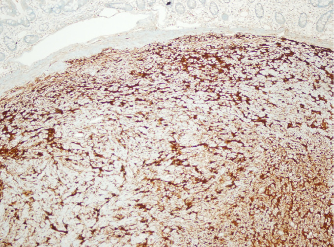
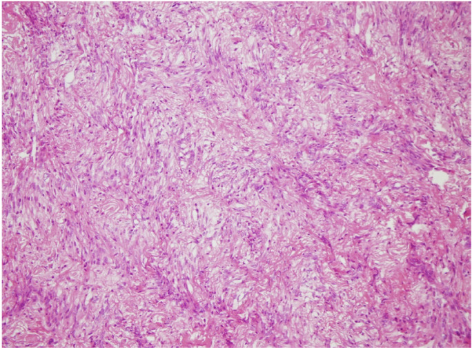
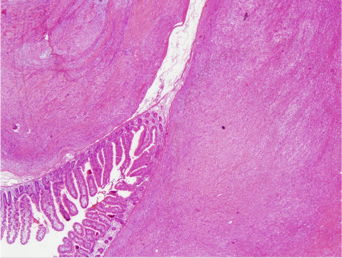
Gastrointestinal schwannomas are benign, slow-growing, rare tumors comprising 2-6% of all mesenchymal tumors of the gastrointestinal tract and 0.2% of all gastric neoplasms. In the gastrointestinal system, schwannomas are mostly observed in the stomach, followed by the colon and rectum. In this case series, we present the clinicopathological results of 9 cases, along with a literature review. A retrospective analysis was conducted on nine patients diagnosed with gastrointestinal schwannoma in a single institution. Tumors were located in the small intestine and stomach, with an average tumor size of 4.6 cm (range: 1.8-8.5 cm). Diagnoses were incidental in most cases, with only four patients presenting symptoms such as epigastric pain and changes in bowel habits. Histopathological characteristics of tumors were studied. Surgical resection with negative margins was performed in 8 cases. Histopathological analysis confirmed schwannomas characterized by solid, homogeneous, spindle-cell structures without cystic changes or necrosis. Immunohistochemically, all tumors were S-100 positive, with variable expression of other markers. Desmin was negative in seven samples. One gastric schwannoma showed focal smooth muscle actin positivity, while others were negative. The Ki-67 index ranged from 0% to 6%, and c-Kit was negative in all cases. DOG-1 expression was examined in four cases, showing focal positivity in small bowel schwannoma and negativity in three gastric schwannomas. Gastrointestinal schwannomas are predominantly benign tumors, more common in women, and typically occur in the sixth decade of life. While imaging and endoscopic techniques help in diagnosis, definitive diagnosis relies on histopathological analysis. Surgical resection remains the gold standard for treatment.

 求助内容:
求助内容: 应助结果提醒方式:


