Andreas Berlin, Lukas Goerdt, Mark E Clark, Liyan Gao, Thomas A Swain, Gerald McGwin, Cynthia Owsley, Kenneth R Sloan, Christine A Curcio
{"title":"Advanced Analysis Tools for Two Wavelength Autofluorescence Imaging of Macular Xanthophyll Carotenoids: ALSTAR2 Baseline.","authors":"Andreas Berlin, Lukas Goerdt, Mark E Clark, Liyan Gao, Thomas A Swain, Gerald McGwin, Cynthia Owsley, Kenneth R Sloan, Christine A Curcio","doi":"10.1167/tvst.14.8.32","DOIUrl":null,"url":null,"abstract":"<p><strong>Purpose: </strong>To allow exploration of xanthophyll carotenoids in vision and age-related macular degeneration progression using two-wavelength autofluorescence imaging for macular pigment optical density (MPOD), we developed tools for automatically centering and classifying the MPOD distribution pattern.</p><p><strong>Methods: </strong>A subset of the ALSTAR2 baseline cohort (NCT04112667) and 44 eyes of adults aged 20 to 30 years with healthy maculas were imaged with optical coherence tomography and two-wavelength autofluorescence (MPOD module, Heidelberg Engineering). Images underwent a quality review. Two custom FIJI plugins centered the MPOD distribution by five algorithms (FOVEA, HILLCLIMB, CENTROID, MAX, CONTOUR). Others automatically classified spatial distributions into four patterns from Obana et al: Peak, Ring, Mixed, and Dip.</p><p><strong>Results: </strong>Of 651 qualifying aged eyes and 44 young eyes, the HILLCLIMB and CONTOUR methods best agreed with a manually determined foveal center. Regarding spatial distribution pattern, 445 aged eyes (68.4%) showed peaks, 118 (18.1%) rings, 41 (6.3%) mixed, and 47 (7.2%) dips. In young eyes, 40 (90%) showed peaks, 1 (2.3%) rings, 3 (6.8%) mixed, and none showed dips. Notably, peaks were significantly (P < 0.001) more prominent in men (74.1%) than women (65.0%) and pseudophakic (72.7%) than phakic (62.9%) eyes.</p><p><strong>Conclusions: </strong>Automatic tools for MPOD centration are reliable and robust. Future studies will use the HILLCLIMB and CONTOUR algorithms.</p><p><strong>Translational relevance: </strong>Automated MPOD pattern assignment suggests that the spatial distribution of MPOD varies with gender, lens status, and possibly age. Our analytic software can be applied to large samples for studies of xanthophyll carotenoid impact on vision and age-related macular degeneration progression.</p>","PeriodicalId":23322,"journal":{"name":"Translational Vision Science & Technology","volume":"14 8","pages":"32"},"PeriodicalIF":2.6000,"publicationDate":"2025-08-01","publicationTypes":"Journal Article","fieldsOfStudy":null,"isOpenAccess":false,"openAccessPdf":"https://www.ncbi.nlm.nih.gov/pmc/articles/PMC12393178/pdf/","citationCount":"0","resultStr":null,"platform":"Semanticscholar","paperid":null,"PeriodicalName":"Translational Vision Science & Technology","FirstCategoryId":"3","ListUrlMain":"https://doi.org/10.1167/tvst.14.8.32","RegionNum":3,"RegionCategory":"医学","ArticlePicture":[],"TitleCN":null,"AbstractTextCN":null,"PMCID":null,"EPubDate":"","PubModel":"","JCR":"Q2","JCRName":"OPHTHALMOLOGY","Score":null,"Total":0}
引用次数: 0
Abstract
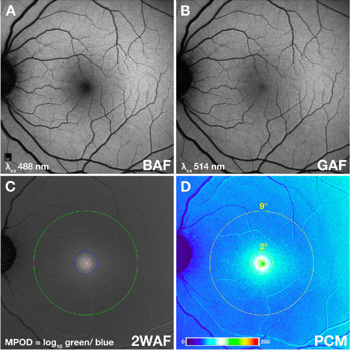
Purpose: To allow exploration of xanthophyll carotenoids in vision and age-related macular degeneration progression using two-wavelength autofluorescence imaging for macular pigment optical density (MPOD), we developed tools for automatically centering and classifying the MPOD distribution pattern.
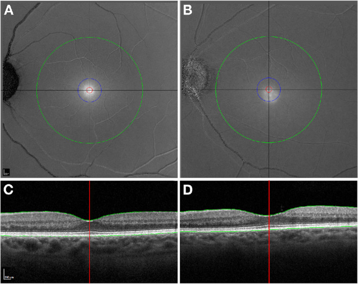
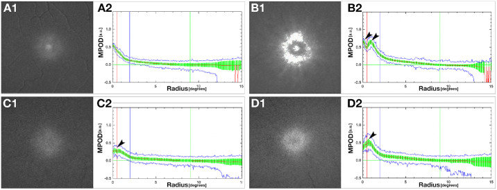
Methods: A subset of the ALSTAR2 baseline cohort (NCT04112667) and 44 eyes of adults aged 20 to 30 years with healthy maculas were imaged with optical coherence tomography and two-wavelength autofluorescence (MPOD module, Heidelberg Engineering). Images underwent a quality review. Two custom FIJI plugins centered the MPOD distribution by five algorithms (FOVEA, HILLCLIMB, CENTROID, MAX, CONTOUR). Others automatically classified spatial distributions into four patterns from Obana et al: Peak, Ring, Mixed, and Dip.
Results: Of 651 qualifying aged eyes and 44 young eyes, the HILLCLIMB and CONTOUR methods best agreed with a manually determined foveal center. Regarding spatial distribution pattern, 445 aged eyes (68.4%) showed peaks, 118 (18.1%) rings, 41 (6.3%) mixed, and 47 (7.2%) dips. In young eyes, 40 (90%) showed peaks, 1 (2.3%) rings, 3 (6.8%) mixed, and none showed dips. Notably, peaks were significantly (P < 0.001) more prominent in men (74.1%) than women (65.0%) and pseudophakic (72.7%) than phakic (62.9%) eyes.
Conclusions: Automatic tools for MPOD centration are reliable and robust. Future studies will use the HILLCLIMB and CONTOUR algorithms.
Translational relevance: Automated MPOD pattern assignment suggests that the spatial distribution of MPOD varies with gender, lens status, and possibly age. Our analytic software can be applied to large samples for studies of xanthophyll carotenoid impact on vision and age-related macular degeneration progression.
期刊介绍:
Translational Vision Science & Technology (TVST), an official journal of the Association for Research in Vision and Ophthalmology (ARVO), an international organization whose purpose is to advance research worldwide into understanding the visual system and preventing, treating and curing its disorders, is an online, open access, peer-reviewed journal emphasizing multidisciplinary research that bridges the gap between basic research and clinical care. A highly qualified and diverse group of Associate Editors and Editorial Board Members is led by Editor-in-Chief Marco Zarbin, MD, PhD, FARVO.
The journal covers a broad spectrum of work, including but not limited to:
Applications of stem cell technology for regenerative medicine,
Development of new animal models of human diseases,
Tissue bioengineering,
Chemical engineering to improve virus-based gene delivery,
Nanotechnology for drug delivery,
Design and synthesis of artificial extracellular matrices,
Development of a true microsurgical operating environment,
Refining data analysis algorithms to improve in vivo imaging technology,
Results of Phase 1 clinical trials,
Reverse translational ("bedside to bench") research.
TVST seeks manuscripts from scientists and clinicians with diverse backgrounds ranging from basic chemistry to ophthalmic surgery that will advance or change the way we understand and/or treat vision-threatening diseases. TVST encourages the use of color, multimedia, hyperlinks, program code and other digital enhancements.

 求助内容:
求助内容: 应助结果提醒方式:


