{"title":"Bronchogenic Cyst with Degeneration of the Adjacent Membranous Portion of the Trachea: A Case Report.","authors":"Takamitsu Hayakawa, Mikako Mitake, Hirohisa Inaba, Mayumi Kobayashi, Yasuhiro Watanabe, Asako Okabe, Kazuhito Funai","doi":"10.70352/scrj.cr.25-0287","DOIUrl":null,"url":null,"abstract":"<p><strong>Introduction: </strong>Bronchogenic cysts are congenital, benign cystic lesions that develop in the mediastinum. Many patients are asymptomatic, and conservative observation is often chosen in clinical practice. However, delayed surgical resection following cyst enlargement and compression of the adjacent membranous portion of the trachea can result in perioperative challenges.</p><p><strong>Case presentation: </strong>We report the case of a 53-year-old woman who had been under observation for 10 years for an asymptomatic mediastinal mass. The mass enlarged gradually and caused persistent cough along with obstructive ventilatory impairment. Chest CT revealed a 5.5 cm mass compressing the membranous trachea, resulting in tracheal stenosis. MRI revealed a homogeneously high T2 signal within the mass, suggesting a simple cystic nature. PET showed no accumulation of fluorodeoxyglucose in the mass, indicating no malignancy. Based on preoperative diagnosis of a bronchogenic cyst, the patient underwent video-assisted thoracoscopic surgery. Tracheal intubation using a double-lumen tube was challenging due to the tracheal stenosis. Moreover, the membranous trachea compressed by the cyst exhibited white degeneration, suggesting thinning and fragility. Intraoperatively, due to firm adhesion to the membranous trachea, a part of the cyst wall was intentionally left in place to avoid tracheal injury. The inner lining of the residual cyst was cauterized to prevent recurrence. Bronchoscopic findings on POD 7 showed that white degeneration of the membranous trachea remained. Histopathological examination revealed ciliated columnar epithelium and cartilage on the cyst wall, confirming the diagnosis of a bronchogenic cyst.</p><p><strong>Conclusions: </strong>Long-term observation of mediastinal bronchogenic cysts can lead to degeneration and thinning of the membranous trachea, increasing the risk of tracheal injury and incomplete resection during surgery. Therefore, the absence of symptoms should not justify delaying surgical intervention. Preoperative assessment for coexisting malignancy and tracheal abnormalities can support surgical decision-making to ensure a safe procedure.</p>","PeriodicalId":22096,"journal":{"name":"Surgical Case Reports","volume":"11 1","pages":""},"PeriodicalIF":0.7000,"publicationDate":"2025-01-01","publicationTypes":"Journal Article","fieldsOfStudy":null,"isOpenAccess":false,"openAccessPdf":"https://www.ncbi.nlm.nih.gov/pmc/articles/PMC12377854/pdf/","citationCount":"0","resultStr":null,"platform":"Semanticscholar","paperid":null,"PeriodicalName":"Surgical Case Reports","FirstCategoryId":"1085","ListUrlMain":"https://doi.org/10.70352/scrj.cr.25-0287","RegionNum":0,"RegionCategory":null,"ArticlePicture":[],"TitleCN":null,"AbstractTextCN":null,"PMCID":null,"EPubDate":"2025/8/20 0:00:00","PubModel":"Epub","JCR":"Q4","JCRName":"SURGERY","Score":null,"Total":0}
引用次数: 0
Abstract
Introduction: Bronchogenic cysts are congenital, benign cystic lesions that develop in the mediastinum. Many patients are asymptomatic, and conservative observation is often chosen in clinical practice. However, delayed surgical resection following cyst enlargement and compression of the adjacent membranous portion of the trachea can result in perioperative challenges.
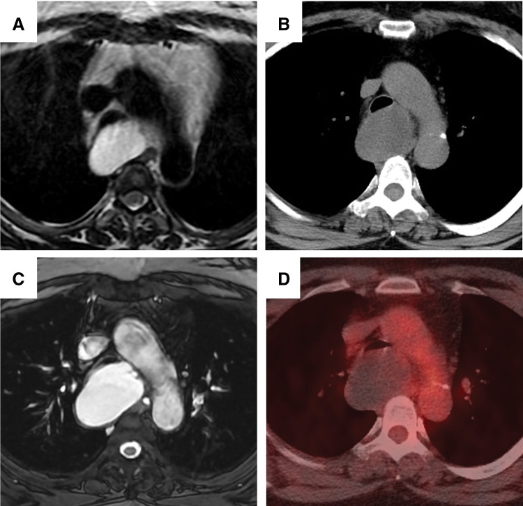
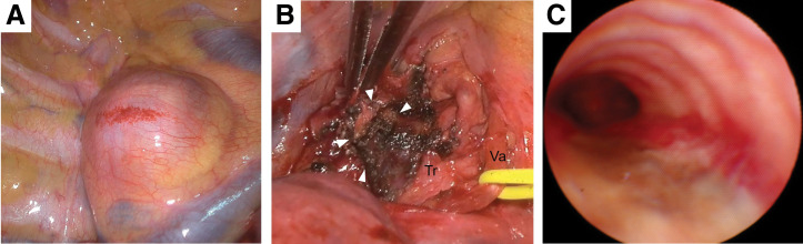
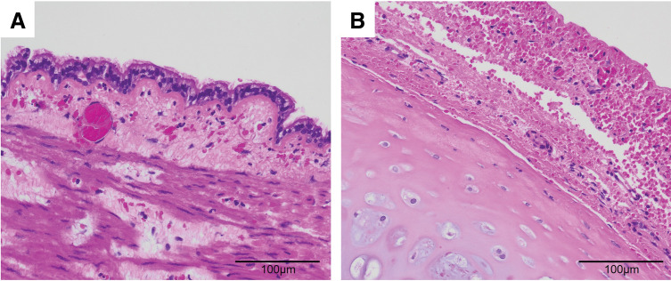
Case presentation: We report the case of a 53-year-old woman who had been under observation for 10 years for an asymptomatic mediastinal mass. The mass enlarged gradually and caused persistent cough along with obstructive ventilatory impairment. Chest CT revealed a 5.5 cm mass compressing the membranous trachea, resulting in tracheal stenosis. MRI revealed a homogeneously high T2 signal within the mass, suggesting a simple cystic nature. PET showed no accumulation of fluorodeoxyglucose in the mass, indicating no malignancy. Based on preoperative diagnosis of a bronchogenic cyst, the patient underwent video-assisted thoracoscopic surgery. Tracheal intubation using a double-lumen tube was challenging due to the tracheal stenosis. Moreover, the membranous trachea compressed by the cyst exhibited white degeneration, suggesting thinning and fragility. Intraoperatively, due to firm adhesion to the membranous trachea, a part of the cyst wall was intentionally left in place to avoid tracheal injury. The inner lining of the residual cyst was cauterized to prevent recurrence. Bronchoscopic findings on POD 7 showed that white degeneration of the membranous trachea remained. Histopathological examination revealed ciliated columnar epithelium and cartilage on the cyst wall, confirming the diagnosis of a bronchogenic cyst.
Conclusions: Long-term observation of mediastinal bronchogenic cysts can lead to degeneration and thinning of the membranous trachea, increasing the risk of tracheal injury and incomplete resection during surgery. Therefore, the absence of symptoms should not justify delaying surgical intervention. Preoperative assessment for coexisting malignancy and tracheal abnormalities can support surgical decision-making to ensure a safe procedure.

 求助内容:
求助内容: 应助结果提醒方式:


