{"title":"Relationship between the prevalence of breast arterial calcifications on mammography and coronary calcifications on Chest CT: A cross-sectional study.","authors":"Laila K Ashkar, Lamia G Jamjoom","doi":"10.15537/smj.2025.46.9.20250316","DOIUrl":null,"url":null,"abstract":"<p><strong>Objectives: </strong>To investigate the relationship between breast arterial calcifications (BAC) detected on mammography and coronary artery calcifications (CAC) identified on chest CT in Saudi women, focusing on prevalence, age-specific patterns, and cardiovascular risk factors.</p><p><strong>Methods: </strong>This cross-sectional study was conducted at a tertiary center in Saudi Arabia using data from hospital electronic medical records of 60 women aged 40-88 years who underwent mammography and chest CT, within the same year during the time period from January 2021 to December 2022. Data on demographics, cardiovascular risk factors, and imaging findings were collected. The association between BAC and CAC was analyzed using chi-square tests and binary logistic regression.</p><p><strong>Results: </strong>The BAC was detected in 33.3% of the participants, while CAC was present in 21.7%. A significant association was observed between the presence of BAC and CAC (<i>p</i>=0.015), with 40.0% of BAC-positive patients showing CAC, compared to only 12.5% of BAC-negative patients. Age was a significant predictor of both BAC and CAC, particularly in the 60-69 age group (<i>p</i>=0.031). Traditional risk factors such as hypertension and diabetes did not show significant predictive value for CAC or BAC.</p><p><strong>Conclusion: </strong>The findings highlight the potential utility of BAC as a non-invasive marker for CAC, particularly in older women. Routine reporting of BAC on mammography could enhance cardiovascular risk stratification in clinical practice.</p>","PeriodicalId":21453,"journal":{"name":"Saudi Medical Journal","volume":"46 9","pages":"1046-1055"},"PeriodicalIF":1.5000,"publicationDate":"2025-09-01","publicationTypes":"Journal Article","fieldsOfStudy":null,"isOpenAccess":false,"openAccessPdf":"https://www.ncbi.nlm.nih.gov/pmc/articles/PMC12441901/pdf/","citationCount":"0","resultStr":null,"platform":"Semanticscholar","paperid":null,"PeriodicalName":"Saudi Medical Journal","FirstCategoryId":"3","ListUrlMain":"https://doi.org/10.15537/smj.2025.46.9.20250316","RegionNum":4,"RegionCategory":"医学","ArticlePicture":[],"TitleCN":null,"AbstractTextCN":null,"PMCID":null,"EPubDate":"","PubModel":"","JCR":"Q2","JCRName":"MEDICINE, GENERAL & INTERNAL","Score":null,"Total":0}
引用次数: 0
Abstract
Objectives: To investigate the relationship between breast arterial calcifications (BAC) detected on mammography and coronary artery calcifications (CAC) identified on chest CT in Saudi women, focusing on prevalence, age-specific patterns, and cardiovascular risk factors.
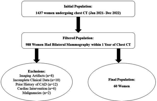
Methods: This cross-sectional study was conducted at a tertiary center in Saudi Arabia using data from hospital electronic medical records of 60 women aged 40-88 years who underwent mammography and chest CT, within the same year during the time period from January 2021 to December 2022. Data on demographics, cardiovascular risk factors, and imaging findings were collected. The association between BAC and CAC was analyzed using chi-square tests and binary logistic regression.
Results: The BAC was detected in 33.3% of the participants, while CAC was present in 21.7%. A significant association was observed between the presence of BAC and CAC (p=0.015), with 40.0% of BAC-positive patients showing CAC, compared to only 12.5% of BAC-negative patients. Age was a significant predictor of both BAC and CAC, particularly in the 60-69 age group (p=0.031). Traditional risk factors such as hypertension and diabetes did not show significant predictive value for CAC or BAC.
Conclusion: The findings highlight the potential utility of BAC as a non-invasive marker for CAC, particularly in older women. Routine reporting of BAC on mammography could enhance cardiovascular risk stratification in clinical practice.
期刊介绍:
The Saudi Medical Journal is a monthly peer-reviewed medical journal. It is an open access journal, with content released under a Creative Commons attribution-noncommercial license.
The journal publishes original research articles, review articles, Systematic Reviews, Case Reports, Brief Communication, Brief Report, Clinical Note, Clinical Image, Editorials, Book Reviews, Correspondence, and Student Corner.
 求助内容:
求助内容: 应助结果提醒方式:


