Henrique Silva, Carlota Rezendes, Pedro Contreiras Pinto
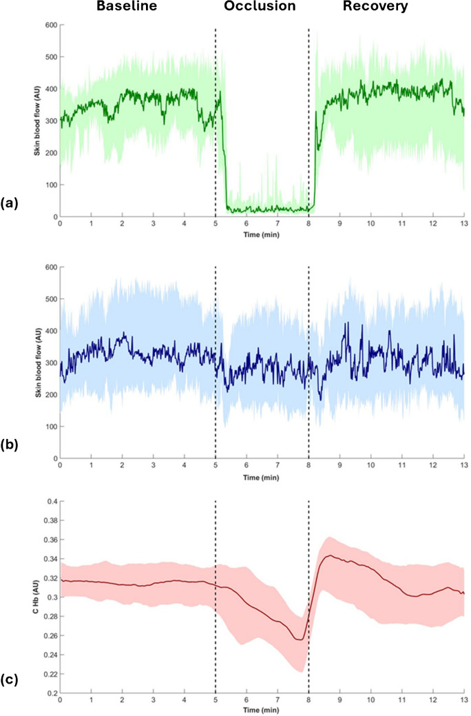
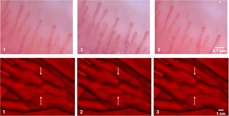
{"title":"Enhancing the quantification of post-occlusive reactive hyperemia: a multimodal optical approach.","authors":"Henrique Silva, Carlota Rezendes, Pedro Contreiras Pinto","doi":"10.1007/s00424-025-03110-7","DOIUrl":null,"url":null,"abstract":"<p><p>Post-occlusive reactive hyperemia (PORH) is a physiological response marked by a transient increase in microvascular perfusion following ischemia. While cutaneous perfusion during PORH has been extensively characterized using optical approaches such as Doppler-based techniques, low-cost alternatives like photoplethysmography (PPG), videocapillaroscopy (VC) and near-infrared reflectance imaging (NIRI) may provide complementary insights into both microvascular and venous dynamics. However, their role in quantifying PORH remains underexplored. This study aimed to evaluate the potential of low-magnification VC and NIRI-based imaging for quantifying perfusion changes during a standardized PORH protocol in healthy subjects, using PPG as a reference. Fourteen participants (21.5 ± 4.2 years) underwent suprasystolic occlusion of a randomly selected upper limb, with simultaneous recordings using PPG and VC at the finger and NIRI at the dorsal hand veins. The protocol included a 5-min baseline, 3-min occlusion (200 mmHg), and 5-min recovery. Skin blood flow was derived from the PPG signal, a hemoglobin index (C<sub>Hb</sub>) was extracted from VC images, and vein width was measured using NIRI. Nonparametric statistics were used for analysis. Arterial occlusion significantly reduced skin blood flow (-95.3%, p < 0.001) and C<sub>Hb</sub> (-8.3%, p = 0.007), with milder contralateral changes. Vein width increased during occlusion (p = 0.003) and returned to baseline during recovery. VC was less sensitive than PPG but reproduced the expected hemodynamic profile. A positive correlation was found between venous dilation during recovery and the decrement velocity of microvascular perfusion during occlusion. VC and NIRI represent accessible and complementary tools for assessing vascular responses during PORH. Their combined application may enhance non-invasive vascular evaluation in both clinical and research settings.</p>","PeriodicalId":19954,"journal":{"name":"Pflugers Archiv : European journal of physiology","volume":" ","pages":"1213-1224"},"PeriodicalIF":2.9000,"publicationDate":"2025-09-01","publicationTypes":"Journal Article","fieldsOfStudy":null,"isOpenAccess":false,"openAccessPdf":"https://www.ncbi.nlm.nih.gov/pmc/articles/PMC12420749/pdf/","citationCount":"0","resultStr":null,"platform":"Semanticscholar","paperid":null,"PeriodicalName":"Pflugers Archiv : European journal of physiology","FirstCategoryId":"3","ListUrlMain":"https://doi.org/10.1007/s00424-025-03110-7","RegionNum":4,"RegionCategory":"医学","ArticlePicture":[],"TitleCN":null,"AbstractTextCN":null,"PMCID":null,"EPubDate":"2025/9/2 0:00:00","PubModel":"Epub","JCR":"Q2","JCRName":"PHYSIOLOGY","Score":null,"Total":0}
引用次数: 0
Abstract
Post-occlusive reactive hyperemia (PORH) is a physiological response marked by a transient increase in microvascular perfusion following ischemia. While cutaneous perfusion during PORH has been extensively characterized using optical approaches such as Doppler-based techniques, low-cost alternatives like photoplethysmography (PPG), videocapillaroscopy (VC) and near-infrared reflectance imaging (NIRI) may provide complementary insights into both microvascular and venous dynamics. However, their role in quantifying PORH remains underexplored. This study aimed to evaluate the potential of low-magnification VC and NIRI-based imaging for quantifying perfusion changes during a standardized PORH protocol in healthy subjects, using PPG as a reference. Fourteen participants (21.5 ± 4.2 years) underwent suprasystolic occlusion of a randomly selected upper limb, with simultaneous recordings using PPG and VC at the finger and NIRI at the dorsal hand veins. The protocol included a 5-min baseline, 3-min occlusion (200 mmHg), and 5-min recovery. Skin blood flow was derived from the PPG signal, a hemoglobin index (CHb) was extracted from VC images, and vein width was measured using NIRI. Nonparametric statistics were used for analysis. Arterial occlusion significantly reduced skin blood flow (-95.3%, p < 0.001) and CHb (-8.3%, p = 0.007), with milder contralateral changes. Vein width increased during occlusion (p = 0.003) and returned to baseline during recovery. VC was less sensitive than PPG but reproduced the expected hemodynamic profile. A positive correlation was found between venous dilation during recovery and the decrement velocity of microvascular perfusion during occlusion. VC and NIRI represent accessible and complementary tools for assessing vascular responses during PORH. Their combined application may enhance non-invasive vascular evaluation in both clinical and research settings.
期刊介绍:
Pflügers Archiv European Journal of Physiology publishes those results of original research that are seen as advancing the physiological sciences, especially those providing mechanistic insights into physiological functions at the molecular and cellular level, and clearly conveying a physiological message. Submissions are encouraged that deal with the evaluation of molecular and cellular mechanisms of disease, ideally resulting in translational research. Purely descriptive papers covering applied physiology or clinical papers will be excluded. Papers on methodological topics will be considered if they contribute to the development of novel tools for further investigation of (patho)physiological mechanisms.
 求助内容:
求助内容: 应助结果提醒方式:


