A versatile miniature two-photon microscope enabling multicolor deep-brain imaging
IF 32.1
1区 生物学
Q1 BIOCHEMICAL RESEARCH METHODS
引用次数: 0
Abstract
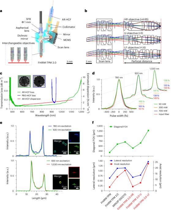
Here we present FHIRM-TPM 3.0, a 2.6 g miniature two-photon microscope capable of multicolor deep-brain imaging in freely behaving mice. The system was integrated with a broadband anti-resonant hollow-core fiber featuring low transmission loss, minimal dispersion from 700 nm to 1,060 nm and high tolerance of laser power. By correcting chromatic and spherical aberrations and optimizing the fluorescence collection aperture, we achieved cortical neuronal imaging at depths exceeding 820 μm and, using a GRIN lens, hippocampal Ca2+ imaging at single dendritic spine resolution. Moreover, we engineered three interchangeable parfocal objectives, allowing for a tenfold scalable field of view up to 1 × 0.8 mm², with lateral resolutions ranging from 0.68 μm to 1.46 μm. By multicolor imaging at excitation wavelengths of 780 nm, 920 nm and 1,030 nm, we investigated mitochondrial and cytosolic Ca2+ activities relative to the deposition of amyloid plaques in the cortex of awake APP/PS1 transgenic mice. Thus, FHIRM-TPM 3.0 provides a versatile imaging system suitable for diverse brain imaging scenarios. FHIRM-TPM 3.0 is a miniature microscope for multicolor two-photon imaging in freely moving mice. In addition to the multicolor imaging abilities achieved with the help of a specially designed optical fiber, the microscope is also compatible with multiple lenses for a choice of field of view and resolution.
多功能微型双光子显微镜,可实现多色脑深部成像。
在这里,我们展示了FHIRM-TPM 3.0,一个2.6 g的微型双光子显微镜,能够在自由行为的小鼠中进行多色脑深部成像。该系统集成了宽带抗谐振空心芯光纤,具有传输损耗低、色散最小(从700 nm到1060 nm)和高激光功率容限的特点。通过校正色差和球差以及优化荧光收集孔径,我们实现了深度超过820 μm的皮质神经元成像,并使用GRIN透镜实现了单树突棘分辨率的海马Ca2+成像。此外,我们设计了三个可互换的共焦物镜,允许10倍可扩展的视场高达1 × 0.8 mm²,横向分辨率范围为0.68 μm至1.46 μm。通过在780 nm、920 nm和1030 nm激发波长下的多色成像,我们研究了清醒APP/PS1转基因小鼠皮层中与淀粉样斑块沉积相关的线粒体和细胞质Ca2+活性。因此,FHIRM-TPM 3.0提供了一个适用于多种脑成像场景的多功能成像系统。
本文章由计算机程序翻译,如有差异,请以英文原文为准。
求助全文
约1分钟内获得全文
求助全文
来源期刊
Nature Methods
生物-生化研究方法
CiteScore
58.70
自引率
1.70%
发文量
326
审稿时长
1 months
期刊介绍:
Nature Methods is a monthly journal that focuses on publishing innovative methods and substantial enhancements to fundamental life sciences research techniques. Geared towards a diverse, interdisciplinary readership of researchers in academia and industry engaged in laboratory work, the journal offers new tools for research and emphasizes the immediate practical significance of the featured work. It publishes primary research papers and reviews recent technical and methodological advancements, with a particular interest in primary methods papers relevant to the biological and biomedical sciences. This includes methods rooted in chemistry with practical applications for studying biological problems.
 求助内容:
求助内容: 应助结果提醒方式:


