Elemental mass spectrometry imaging of biomolecules using metal-conjugated probes
IF 51.7
1区 化学
Q1 CHEMISTRY, MULTIDISCIPLINARY
引用次数: 0
Abstract
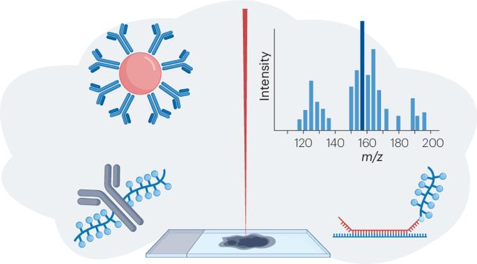
In situ imaging of proteins, RNA, immune cells and other biomolecules is necessary to determine their function, interactions and roles in disease pathology. Increasingly, this is achieved via metal-conjugated probes in conjunction with elemental mass spectrometry imaging (MSI). This targeted technique is capable of simultaneously imaging up to 40 analytes, in comparison to the traditional bioimaging techniques that use fluorescent or chromogenic reagents that are typically restricted to less than four analytes without complex sample handling and analysis workflows. These analyses, however, are not straightforward, with a number of factors that require optimization. They require the use of probes specific to the target biomolecules, which are conjugated with analytes detectable by elemental MSI. Here, we summarize the MSI technology, the types of biological probes used for identification, and the forms of metal analytes used. We provide examples of their application including understanding cancer cell heterogeneity to direct clinical trials, which may impact clinical diagnostics and personalized medicine. We conclude with future perspectives on the potential of the technique and what is required to meet it. Elemental mass spectrometry imaging of biomolecules provides detailed knowledge of their abundance and location within tissue samples. This Review highlights the analytical instrumentation and strategies used to bring this technique from a research tool to clinical studies.
使用金属共轭探针的生物分子元素质谱成像。
蛋白质、RNA、免疫细胞和其他生物分子的原位成像对于确定它们在疾病病理中的功能、相互作用和作用是必要的。越来越多地,这是通过金属共轭探针结合元素质谱成像(MSI)来实现的。与传统的生物成像技术相比,这种靶向技术能够同时成像多达40种分析物,而传统的生物成像技术使用荧光或显色试剂,通常限于少于4种分析物,没有复杂的样品处理和分析工作流程。然而,这些分析并不简单,有许多因素需要优化。它们需要使用特定于目标生物分子的探针,这些探针与元素MSI可检测的分析物偶联。在这里,我们总结了MSI技术,用于鉴定的生物探针类型,以及所使用的金属分析物的形式。我们提供了它们应用的例子,包括了解癌细胞异质性到直接临床试验,这可能会影响临床诊断和个性化医疗。最后,我们对该技术的潜力和满足它所需要的未来前景进行了展望。
本文章由计算机程序翻译,如有差异,请以英文原文为准。
求助全文
约1分钟内获得全文
求助全文
来源期刊
Nature reviews. Chemistry
Chemical Engineering-General Chemical Engineering
CiteScore
52.80
自引率
0.80%
发文量
88
期刊介绍:
Nature Reviews Chemistry is an online-only journal that publishes Reviews, Perspectives, and Comments on various disciplines within chemistry. The Reviews aim to offer balanced and objective analyses of selected topics, providing clear descriptions of relevant scientific literature. The content is designed to be accessible to recent graduates in any chemistry-related discipline while also offering insights for principal investigators and industry-based research scientists. Additionally, Reviews should provide the authors' perspectives on future directions and opinions regarding the major challenges faced by researchers in the field.
 求助内容:
求助内容: 应助结果提醒方式:


