Christopher Foster, Ryan Tran, Khushi Grover, Abdullah Salama, Courtney K Rowe
{"title":"Development of an Ex Vivo Platform to Model Urethral Healing.","authors":"Christopher Foster, Ryan Tran, Khushi Grover, Abdullah Salama, Courtney K Rowe","doi":"10.3390/mps8040096","DOIUrl":null,"url":null,"abstract":"<p><strong>Background: </strong>Urethral strictures impact millions, causing significant morbidity and millions in healthcare costs. Testing new interventions is limited by the lack of inexpensive urethral healing models. We developed an ex vivo model of early urethral wound healing using explanted rabbit urethral tissue. This was used to test the impact of six growth factors (GFs).</p><p><strong>Methods: </strong>The rabbit urethra was detubularized by cutting it between the corpora cavernosa, and then it was stitched flat using a custom 3D-printed platform. The tissue was carefully scratched to produce a visible wound, and the specimens were placed in media containing growth factors at 100 ng/mL and 10 ng/mL. Images were taken at 0, 24, 48, 72, and 96 h, and the wound area was measured by blinded reviewers to determine the rate of wound contraction.</p><p><strong>Results: </strong>Specimens with IGF at 100 ng/mL showed a statistically significant difference in wound contraction when compared to those with GF-free control medium, showing that IGF-1 supports early urethral epithelization and may improve healing.</p><p><strong>Conclusions: </strong>The developed protocol provides a simple explant platform that can be used to investigate methods of enhancing early phases of urethral healing or used to investigate other areas of urethral health, including drug delivery, infection, and mechanical properties.</p>","PeriodicalId":18715,"journal":{"name":"Methods and Protocols","volume":"8 4","pages":""},"PeriodicalIF":2.0000,"publicationDate":"2025-08-15","publicationTypes":"Journal Article","fieldsOfStudy":null,"isOpenAccess":false,"openAccessPdf":"https://www.ncbi.nlm.nih.gov/pmc/articles/PMC12388648/pdf/","citationCount":"0","resultStr":null,"platform":"Semanticscholar","paperid":null,"PeriodicalName":"Methods and Protocols","FirstCategoryId":"1085","ListUrlMain":"https://doi.org/10.3390/mps8040096","RegionNum":0,"RegionCategory":null,"ArticlePicture":[],"TitleCN":null,"AbstractTextCN":null,"PMCID":null,"EPubDate":"","PubModel":"","JCR":"Q3","JCRName":"BIOCHEMICAL RESEARCH METHODS","Score":null,"Total":0}
引用次数: 0
Abstract
Background: Urethral strictures impact millions, causing significant morbidity and millions in healthcare costs. Testing new interventions is limited by the lack of inexpensive urethral healing models. We developed an ex vivo model of early urethral wound healing using explanted rabbit urethral tissue. This was used to test the impact of six growth factors (GFs).
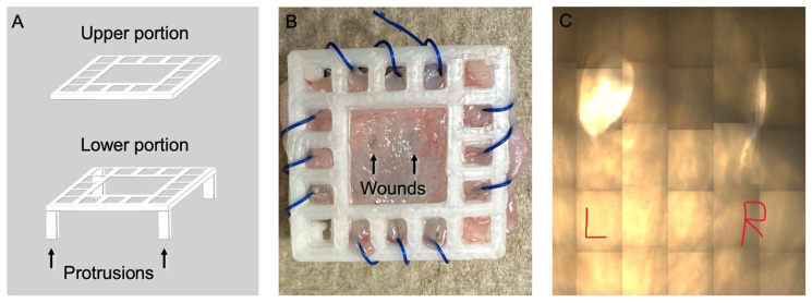
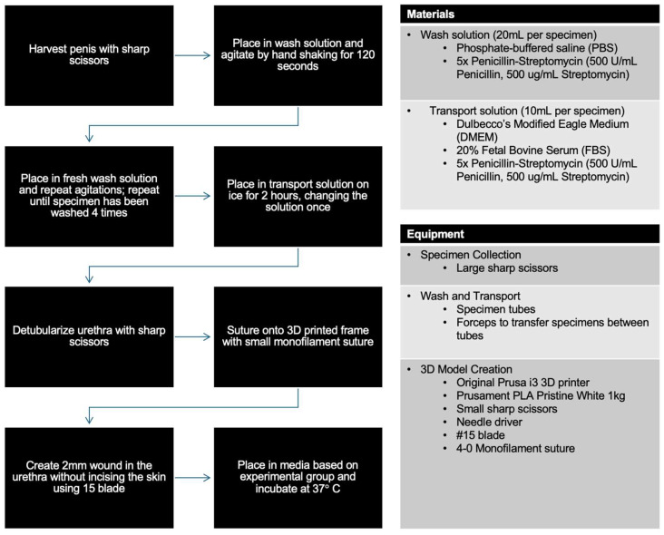
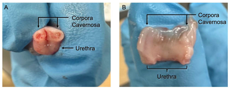
Methods: The rabbit urethra was detubularized by cutting it between the corpora cavernosa, and then it was stitched flat using a custom 3D-printed platform. The tissue was carefully scratched to produce a visible wound, and the specimens were placed in media containing growth factors at 100 ng/mL and 10 ng/mL. Images were taken at 0, 24, 48, 72, and 96 h, and the wound area was measured by blinded reviewers to determine the rate of wound contraction.
Results: Specimens with IGF at 100 ng/mL showed a statistically significant difference in wound contraction when compared to those with GF-free control medium, showing that IGF-1 supports early urethral epithelization and may improve healing.
Conclusions: The developed protocol provides a simple explant platform that can be used to investigate methods of enhancing early phases of urethral healing or used to investigate other areas of urethral health, including drug delivery, infection, and mechanical properties.

 求助内容:
求助内容: 应助结果提醒方式:


