Artificial Intelligence Analysis of Chest Radiographs for Predicting Major Adverse Events in Patients Visiting the Emergency Department With Acute Cardiopulmonary Symptoms.
IF 5.3 2区 医学Q1 RADIOLOGY, NUCLEAR MEDICINE & MEDICAL IMAGING
Chanyoung Rhee, Ki Jeong Hong, Ki Hong Kim, Jin Mo Goo, Eui Jin Hwang
{"title":"Artificial Intelligence Analysis of Chest Radiographs for Predicting Major Adverse Events in Patients Visiting the Emergency Department With Acute Cardiopulmonary Symptoms.","authors":"Chanyoung Rhee, Ki Jeong Hong, Ki Hong Kim, Jin Mo Goo, Eui Jin Hwang","doi":"10.3348/kjr.2025.0237","DOIUrl":null,"url":null,"abstract":"<p><strong>Objective: </strong>In this study, we investigated whether artificial intelligence (AI) analysis of chest radiographs (CXRs) can predict major adverse clinical events in patients visiting the emergency department (ED) with acute cardiopulmonary symptoms.</p><p><strong>Materials and methods: </strong>This secondary analysis of a previous clinical trial included patients who visited the ED with symptoms suggestive of acute cardiopulmonary disease and underwent chest radiography between June 2020 and December 2021. All patients underwent triage upon arrival at ED according to the Korean Triage and Acuity Scale (KTAS). The CXRs were retrospectively analyzed using a commercial AI (Lunit INSIGHT CXR, version 3.1.4.1) capable of detecting seven abnormalities on a single frontal CXR. The predictive performance of the AI analysis for major adverse cardiopulmonary events (any among hospitalization, ED revisits, and death in the ED due to acute cardiopulmonary disease) was compared with that of the KTAS using the area under the receiver operating characteristic curve (AUC). Multivariable (the AI analysis result and KTAS level) logistic regression analysis was conducted to investigate whether the AI analysis result was an independent predictor of the events and whether the combination of the AI analysis and KTAS has additional merit.</p><p><strong>Results: </strong>Among 3576 patients (1966 males; mean age, 64 years), 1148 (32.1%) experienced major adverse cardiopulmonary events. AI analysis of CXRs outperformed the KTAS in predicting these events (AUC, 0.795 vs. 0.610; <i>P</i> < 0.001). The AI analysis result was an independent predictor of these events after adjusting for the KTAS level (adjusted odd ratios of 1.032 and 6.913 for every 1% increase and ≥15%, respectively, in the AI probability score; <i>P</i> < 0.001). The combination of the AI analysis and KTAS showed an AUC that was higher than that of the KTAS alone (0.799; <i>P</i> < 0.001) and in-par with that of the AI analysis only (<i>P</i> = 0.187).</p><p><strong>Conclusion: </strong>AI analysis of CXRs showed greater accuracy than the KTAS did in predicting major adverse cardiopulmonary events in patients visiting the ED with acute cardiopulmonary symptoms. AI analysis may enhance the efficacy of patient triage in the ED.</p>","PeriodicalId":17881,"journal":{"name":"Korean Journal of Radiology","volume":"26 9","pages":"877-887"},"PeriodicalIF":5.3000,"publicationDate":"2025-09-01","publicationTypes":"Journal Article","fieldsOfStudy":null,"isOpenAccess":false,"openAccessPdf":"https://www.ncbi.nlm.nih.gov/pmc/articles/PMC12394822/pdf/","citationCount":"0","resultStr":null,"platform":"Semanticscholar","paperid":null,"PeriodicalName":"Korean Journal of Radiology","FirstCategoryId":"3","ListUrlMain":"https://doi.org/10.3348/kjr.2025.0237","RegionNum":2,"RegionCategory":"医学","ArticlePicture":[],"TitleCN":null,"AbstractTextCN":null,"PMCID":null,"EPubDate":"","PubModel":"","JCR":"Q1","JCRName":"RADIOLOGY, NUCLEAR MEDICINE & MEDICAL IMAGING","Score":null,"Total":0}
引用次数: 0
Abstract
Objective: In this study, we investigated whether artificial intelligence (AI) analysis of chest radiographs (CXRs) can predict major adverse clinical events in patients visiting the emergency department (ED) with acute cardiopulmonary symptoms.
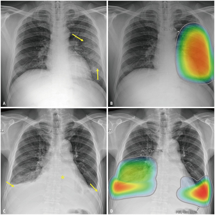
Materials and methods: This secondary analysis of a previous clinical trial included patients who visited the ED with symptoms suggestive of acute cardiopulmonary disease and underwent chest radiography between June 2020 and December 2021. All patients underwent triage upon arrival at ED according to the Korean Triage and Acuity Scale (KTAS). The CXRs were retrospectively analyzed using a commercial AI (Lunit INSIGHT CXR, version 3.1.4.1) capable of detecting seven abnormalities on a single frontal CXR. The predictive performance of the AI analysis for major adverse cardiopulmonary events (any among hospitalization, ED revisits, and death in the ED due to acute cardiopulmonary disease) was compared with that of the KTAS using the area under the receiver operating characteristic curve (AUC). Multivariable (the AI analysis result and KTAS level) logistic regression analysis was conducted to investigate whether the AI analysis result was an independent predictor of the events and whether the combination of the AI analysis and KTAS has additional merit.
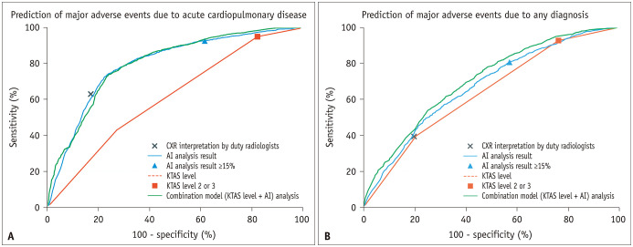
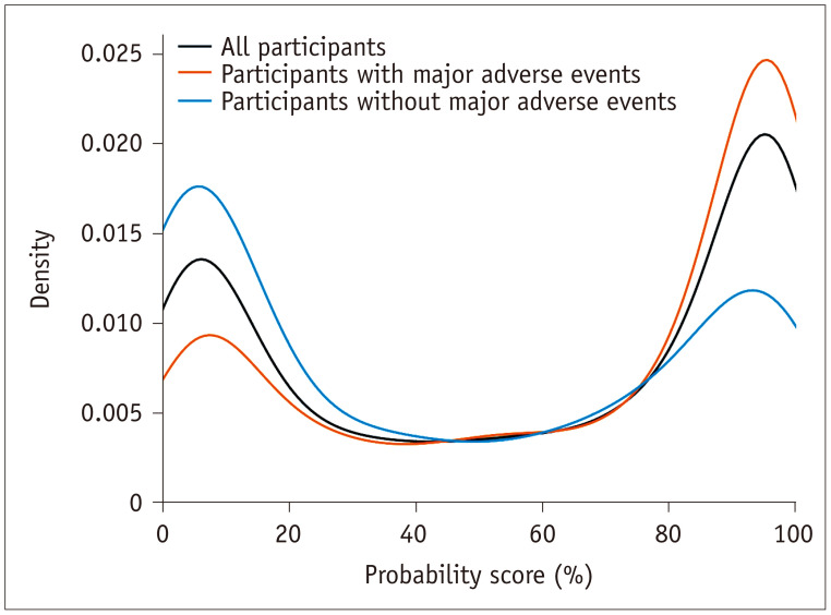
Results: Among 3576 patients (1966 males; mean age, 64 years), 1148 (32.1%) experienced major adverse cardiopulmonary events. AI analysis of CXRs outperformed the KTAS in predicting these events (AUC, 0.795 vs. 0.610; P < 0.001). The AI analysis result was an independent predictor of these events after adjusting for the KTAS level (adjusted odd ratios of 1.032 and 6.913 for every 1% increase and ≥15%, respectively, in the AI probability score; P < 0.001). The combination of the AI analysis and KTAS showed an AUC that was higher than that of the KTAS alone (0.799; P < 0.001) and in-par with that of the AI analysis only (P = 0.187).
Conclusion: AI analysis of CXRs showed greater accuracy than the KTAS did in predicting major adverse cardiopulmonary events in patients visiting the ED with acute cardiopulmonary symptoms. AI analysis may enhance the efficacy of patient triage in the ED.
期刊介绍:
The inaugural issue of the Korean J Radiol came out in March 2000. Our journal aims to produce and propagate knowledge on radiologic imaging and related sciences.
A unique feature of the articles published in the Journal will be their reflection of global trends in radiology combined with an East-Asian perspective. Geographic differences in disease prevalence will be reflected in the contents of papers, and this will serve to enrich our body of knowledge.
World''s outstanding radiologists from many countries are serving as editorial board of our journal.

 求助内容:
求助内容: 应助结果提醒方式:


