{"title":"Dynamics of color vision recovery in Vogt-Koyanagi-Harada disease: a longitudinal study using cone contrast test and adaptive optics imaging.","authors":"Tomoko Nakamura, Shinya Abe, Hitoshi Yamazaki, Toshihiko Oiwake, Atsushi Hayashi","doi":"10.1186/s12348-025-00523-4","DOIUrl":null,"url":null,"abstract":"<p><strong>Background: </strong>Vogt-Koyanagi-Harada (VKH) disease affects visual function, but the recovery process of color vision remains incompletely understood. This study aimed to assess color vision recovery in VKH using cone contrast testing and explore its relationship with cone cell density measured using adaptive optics imaging.</p><p><strong>Methods: </strong>Twenty-two eyes of 11 patients with VKH were evaluated at baseline (serous retinal detachment resolution) and at 3, 6, and 12 months post-treatment. Cone contrast scores and cone cell density were measured using the ColorDx<sup>®</sup> CCT-HD™ system and rtx1™ adaptive optics fundus camera, respectively. Pre-treatment optical coherence tomography (OCT) was used to analyze the cystoid spaces.</p><p><strong>Results: </strong>Color vision defects observed at baseline-especially in S-cones-significantly improved by 6 months for all cone types. Cone contrast scores correlated significantly with cone cell density (ρ = 0.63-0.66, p < 0.0001). Eyes with cystoid spaces on pre-treatment OCT showed lower cone contrast scores and cone density. S-cones demonstrated greater vulnerability and slower recovery than L- and M-cones.</p><p><strong>Conclusion: </strong>This study emphasizes the importance of comprehensive visual function assessment, including color vision evaluation, in VKH patients. Cone contrast testing captures qualitative aspects of visual function not reflected in standard acuity tests. The combination of cone contrast testing and adaptive optics imaging provides insights into the structure-function relationship in color vision recovery.</p>","PeriodicalId":16600,"journal":{"name":"Journal of Ophthalmic Inflammation and Infection","volume":"15 1","pages":"64"},"PeriodicalIF":2.3000,"publicationDate":"2025-08-25","publicationTypes":"Journal Article","fieldsOfStudy":null,"isOpenAccess":false,"openAccessPdf":"https://www.ncbi.nlm.nih.gov/pmc/articles/PMC12378258/pdf/","citationCount":"0","resultStr":null,"platform":"Semanticscholar","paperid":null,"PeriodicalName":"Journal of Ophthalmic Inflammation and Infection","FirstCategoryId":"1085","ListUrlMain":"https://doi.org/10.1186/s12348-025-00523-4","RegionNum":0,"RegionCategory":null,"ArticlePicture":[],"TitleCN":null,"AbstractTextCN":null,"PMCID":null,"EPubDate":"","PubModel":"","JCR":"Q1","JCRName":"OPHTHALMOLOGY","Score":null,"Total":0}
引用次数: 0
Abstract
Background: Vogt-Koyanagi-Harada (VKH) disease affects visual function, but the recovery process of color vision remains incompletely understood. This study aimed to assess color vision recovery in VKH using cone contrast testing and explore its relationship with cone cell density measured using adaptive optics imaging.
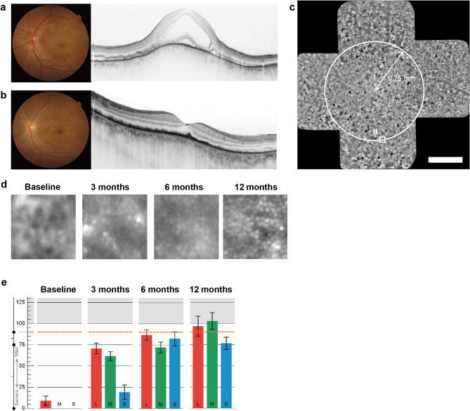
Methods: Twenty-two eyes of 11 patients with VKH were evaluated at baseline (serous retinal detachment resolution) and at 3, 6, and 12 months post-treatment. Cone contrast scores and cone cell density were measured using the ColorDx® CCT-HD™ system and rtx1™ adaptive optics fundus camera, respectively. Pre-treatment optical coherence tomography (OCT) was used to analyze the cystoid spaces.
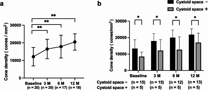
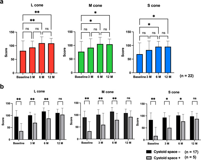
Results: Color vision defects observed at baseline-especially in S-cones-significantly improved by 6 months for all cone types. Cone contrast scores correlated significantly with cone cell density (ρ = 0.63-0.66, p < 0.0001). Eyes with cystoid spaces on pre-treatment OCT showed lower cone contrast scores and cone density. S-cones demonstrated greater vulnerability and slower recovery than L- and M-cones.
Conclusion: This study emphasizes the importance of comprehensive visual function assessment, including color vision evaluation, in VKH patients. Cone contrast testing captures qualitative aspects of visual function not reflected in standard acuity tests. The combination of cone contrast testing and adaptive optics imaging provides insights into the structure-function relationship in color vision recovery.
 求助内容:
求助内容: 应助结果提醒方式:


