Zhiwang Guo, Tao Wu, Xiaolei Chen, Huimin Chang, Bing Hou
{"title":"Intraventricular metastasis with malignant progression of an intraspinal solitary fibrous tumor: a case report and review of the literature.","authors":"Zhiwang Guo, Tao Wu, Xiaolei Chen, Huimin Chang, Bing Hou","doi":"10.1186/s13256-025-05518-2","DOIUrl":null,"url":null,"abstract":"<p><strong>Background: </strong>Solitary fibrous tumors are rare central nervous system neoplasms with high rates of local recurrence and distant metastasis. To date, no instances of metastatic dissemination from the spine to the ventricle with malignant progression have been documented.</p><p><strong>Case presentation: </strong>A 32-year-old male patient of Chinese ethnicity was diagnosed with a grade 2 intraspinal solitary fibrous tumor, demonstrating metastatic spread to the trigone region of the lateral ventricle. Despite cystic degeneration, the solid component of the metastatic lesion exhibited signal intensity similar to that of the primary tumor on imaging. A piecemeal gross total resection was achieved, and postoperative immunohistochemical analysis confirmed that the metastatic lesion was categorized as grade 3, with an increase in the Ki-67 proliferation index from 20% to 30%. Both the primary and metastatic tumors exhibited negative expression of CD34. Although radiotherapy was considered, consensus on its use was not reached, and the patient succumbed to tumor progression 17 months later.</p><p><strong>Conclusion: </strong>This case underscores the importance of recognizing the potential for malignant transformation and intracranial metastasis in intraspinal solitary fibrous tumors, highlighting the need for vigilant monitoring and possible aggressive treatment strategies.</p>","PeriodicalId":16236,"journal":{"name":"Journal of Medical Case Reports","volume":"19 1","pages":"432"},"PeriodicalIF":0.8000,"publicationDate":"2025-08-30","publicationTypes":"Journal Article","fieldsOfStudy":null,"isOpenAccess":false,"openAccessPdf":"https://www.ncbi.nlm.nih.gov/pmc/articles/PMC12398998/pdf/","citationCount":"0","resultStr":null,"platform":"Semanticscholar","paperid":null,"PeriodicalName":"Journal of Medical Case Reports","FirstCategoryId":"1085","ListUrlMain":"https://doi.org/10.1186/s13256-025-05518-2","RegionNum":0,"RegionCategory":null,"ArticlePicture":[],"TitleCN":null,"AbstractTextCN":null,"PMCID":null,"EPubDate":"","PubModel":"","JCR":"Q3","JCRName":"MEDICINE, GENERAL & INTERNAL","Score":null,"Total":0}
引用次数: 0
Abstract
Background: Solitary fibrous tumors are rare central nervous system neoplasms with high rates of local recurrence and distant metastasis. To date, no instances of metastatic dissemination from the spine to the ventricle with malignant progression have been documented.
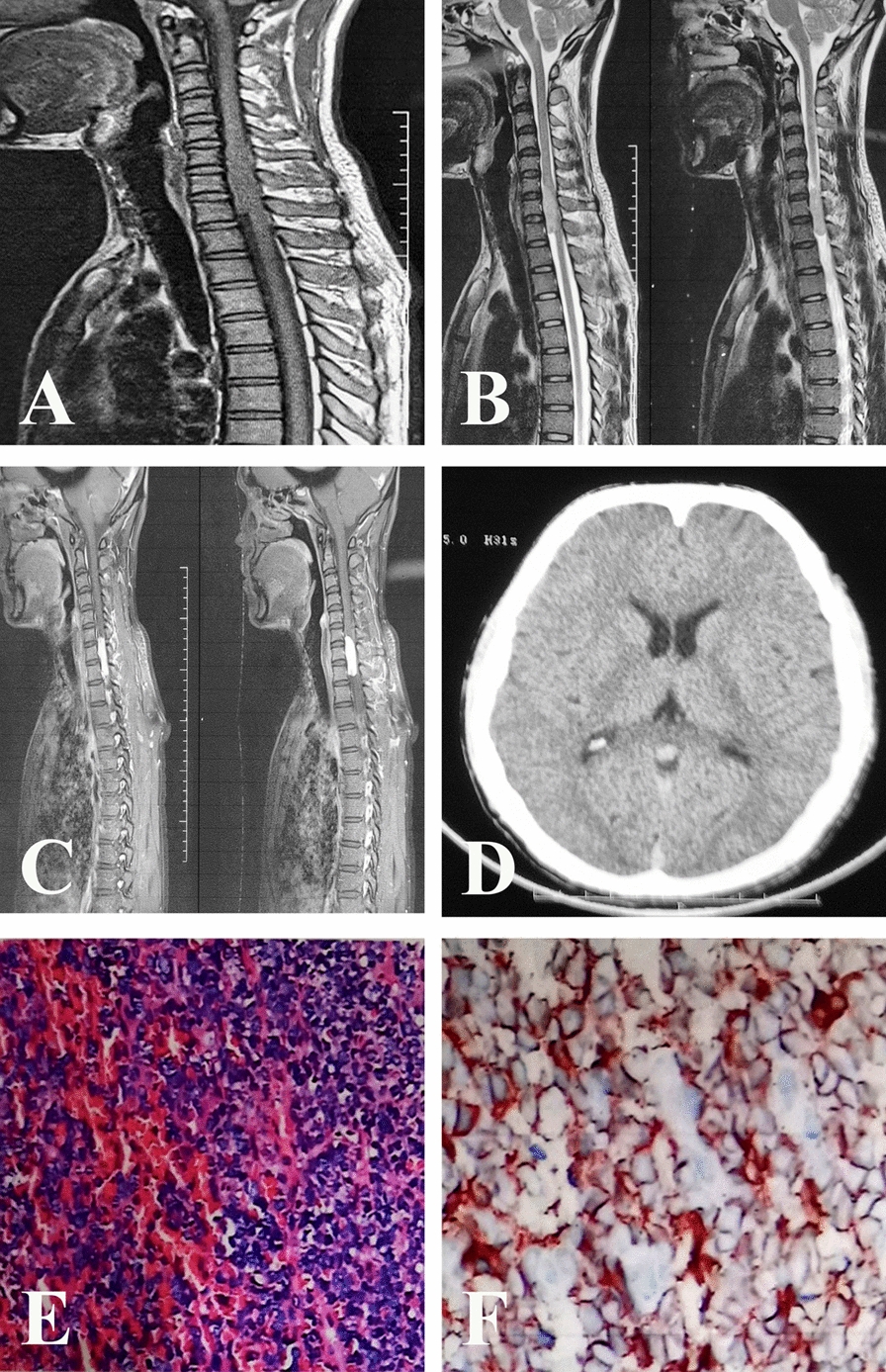
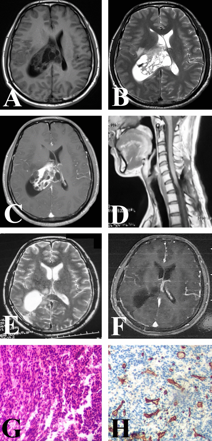
Case presentation: A 32-year-old male patient of Chinese ethnicity was diagnosed with a grade 2 intraspinal solitary fibrous tumor, demonstrating metastatic spread to the trigone region of the lateral ventricle. Despite cystic degeneration, the solid component of the metastatic lesion exhibited signal intensity similar to that of the primary tumor on imaging. A piecemeal gross total resection was achieved, and postoperative immunohistochemical analysis confirmed that the metastatic lesion was categorized as grade 3, with an increase in the Ki-67 proliferation index from 20% to 30%. Both the primary and metastatic tumors exhibited negative expression of CD34. Although radiotherapy was considered, consensus on its use was not reached, and the patient succumbed to tumor progression 17 months later.
Conclusion: This case underscores the importance of recognizing the potential for malignant transformation and intracranial metastasis in intraspinal solitary fibrous tumors, highlighting the need for vigilant monitoring and possible aggressive treatment strategies.
期刊介绍:
JMCR is an open access, peer-reviewed online journal that will consider any original case report that expands the field of general medical knowledge. Reports should show one of the following: 1. Unreported or unusual side effects or adverse interactions involving medications 2. Unexpected or unusual presentations of a disease 3. New associations or variations in disease processes 4. Presentations, diagnoses and/or management of new and emerging diseases 5. An unexpected association between diseases or symptoms 6. An unexpected event in the course of observing or treating a patient 7. Findings that shed new light on the possible pathogenesis of a disease or an adverse effect

 求助内容:
求助内容: 应助结果提醒方式:


