{"title":"Investigation of Trunk and Pelvis Muscle Activity during Sprinting using T2-Weighted Magnetic Resonance Imaging.","authors":"Takaya Yoshimoto, Yoshihiro Chiba, Hayato Ohnuma, Norihide Sugisaki","doi":"10.5114/jhk/197315","DOIUrl":null,"url":null,"abstract":"<p><p>There are few studies that clarify the level of muscle activity in the trunk and pelvis muscles during sprinting. This study aimed to investigate muscle activity in the trunk and pelvis muscles during sprinting using T2-weighted magnetic resonance imaging (MRI). The pre- and post-test designs were employed by measuring trunk and pelvis muscle activity using T2-weighted MRI before and after 60-m round-trip sprints. Ten male sprinters (N = 10, age, 23.3 ± 6.7 years; body height, 175.1 ± 3.6 cm; body mass, 66.8 ± 4.3 kg; 100-m personal record, 11.18 ± 0.48 s, means ± standard deviations [SDs]) performed three sets of three 60-m round-trip sprints. Before and after the round-trip sprints, 3T MRI scans were performed to obtain the T2 values of the trunk and pelvis muscles. After the 60-m roundtrip sprints, the T2 values of lateral abdominal, psoas major, erector spinae, gluteus maximus, gluteus medius, rectus femoris, tensor fasciae latae, sartorius and pectineus muscles increased significantly. There were intermuscular differences in the rate of change of T2 values before and after the 60-m round-trip sprints, with significantly higher levels of muscle activity in lateral abdominals, psoas major, erector spinae, gluteus maximus, and pectineus. In sprinting, the trunk and pelvis muscles were found to be specifically activated.</p>","PeriodicalId":16055,"journal":{"name":"Journal of Human Kinetics","volume":"98 ","pages":"195-204"},"PeriodicalIF":2.8000,"publicationDate":"2025-05-29","publicationTypes":"Journal Article","fieldsOfStudy":null,"isOpenAccess":false,"openAccessPdf":"https://www.ncbi.nlm.nih.gov/pmc/articles/PMC12360926/pdf/","citationCount":"0","resultStr":null,"platform":"Semanticscholar","paperid":null,"PeriodicalName":"Journal of Human Kinetics","FirstCategoryId":"3","ListUrlMain":"https://doi.org/10.5114/jhk/197315","RegionNum":3,"RegionCategory":"医学","ArticlePicture":[],"TitleCN":null,"AbstractTextCN":null,"PMCID":null,"EPubDate":"2025/7/1 0:00:00","PubModel":"eCollection","JCR":"Q2","JCRName":"SPORT SCIENCES","Score":null,"Total":0}
引用次数: 0
Abstract
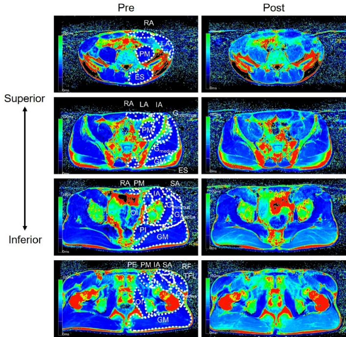
There are few studies that clarify the level of muscle activity in the trunk and pelvis muscles during sprinting. This study aimed to investigate muscle activity in the trunk and pelvis muscles during sprinting using T2-weighted magnetic resonance imaging (MRI). The pre- and post-test designs were employed by measuring trunk and pelvis muscle activity using T2-weighted MRI before and after 60-m round-trip sprints. Ten male sprinters (N = 10, age, 23.3 ± 6.7 years; body height, 175.1 ± 3.6 cm; body mass, 66.8 ± 4.3 kg; 100-m personal record, 11.18 ± 0.48 s, means ± standard deviations [SDs]) performed three sets of three 60-m round-trip sprints. Before and after the round-trip sprints, 3T MRI scans were performed to obtain the T2 values of the trunk and pelvis muscles. After the 60-m roundtrip sprints, the T2 values of lateral abdominal, psoas major, erector spinae, gluteus maximus, gluteus medius, rectus femoris, tensor fasciae latae, sartorius and pectineus muscles increased significantly. There were intermuscular differences in the rate of change of T2 values before and after the 60-m round-trip sprints, with significantly higher levels of muscle activity in lateral abdominals, psoas major, erector spinae, gluteus maximus, and pectineus. In sprinting, the trunk and pelvis muscles were found to be specifically activated.
期刊介绍:
The Journal of Human Kinetics is an open access interdisciplinary periodical offering the latest research in the science of human movement studies. This comprehensive professional journal features articles and research notes encompassing such topic areas as: Kinesiology, Exercise Physiology and Nutrition, Sports Training and Behavioural Sciences in Sport, but especially considering elite and competitive aspects of sport.
The journal publishes original papers, invited reviews, short communications and letters to the Editors. Manuscripts submitted to the journal must contain novel data on theoretical or experimental research or on practical applications in the field of sport sciences.
The Journal of Human Kinetics is published in March, June, September and December.
We encourage scientists from around the world to submit their papers to our periodical.
 求助内容:
求助内容: 应助结果提醒方式:


