Hunaina Shahab, Jad Kassem, Ali Yildiz, Adam H Jacobi
{"title":"Thrombosed saphenous vein graft aneurysm mimicking left atrial appendage mass: A rare complication of coronary artery bypass graft.","authors":"Hunaina Shahab, Jad Kassem, Ali Yildiz, Adam H Jacobi","doi":"10.25259/JCIS_45_2025","DOIUrl":null,"url":null,"abstract":"<p><p>Saphenous vein graft aneurysm (SVGA) is a rare but potentially life-threatening complication of coronary artery bypass grafting (CABG). Its incidence is likely underreported due to asymptomatic cases and undiagnosed acute events. While SVGAs are more commonly associated with right atrial compression, presentation as a left atrial mass is rare. We present the case of an 85-year-old man with a history of CABG, who was incidentally found to have a left atrial appendage (LAA) density on a computed tomography (CT) chest, abdomen, and pelvis performed for unrelated symptoms of back pain and constipation. The density was initially suspected to be an LAA thrombus. However, a dedicated cardiac CT with delayed-phase imaging revealed a largely thrombosed aneurysmal saphenous vein graft to the obtuse marginal artery, which indented the LAA, mimicking an intracardiac mass. This case underscores the critical role of multimodality imaging, particularly cardiac CT, in differentiating vascular aneurysms from true intracardiac masses. Given the patient's asymptomatic status, conservative management with close follow-up was pursued. This case adds to the limited literature on SVGAs mimicking left atrial pathology and highlights the importance of recognizing this rare entity to avoid unnecessary interventions. It also emphasizes the evolving role of cardiac CT as a noninvasive, high-yield diagnostic tool for complex post-CABG anatomical assessments.</p>","PeriodicalId":15512,"journal":{"name":"Journal of Clinical Imaging Science","volume":"15 ","pages":"26"},"PeriodicalIF":1.3000,"publicationDate":"2025-07-16","publicationTypes":"Journal Article","fieldsOfStudy":null,"isOpenAccess":false,"openAccessPdf":"https://www.ncbi.nlm.nih.gov/pmc/articles/PMC12361656/pdf/","citationCount":"0","resultStr":null,"platform":"Semanticscholar","paperid":null,"PeriodicalName":"Journal of Clinical Imaging Science","FirstCategoryId":"1085","ListUrlMain":"https://doi.org/10.25259/JCIS_45_2025","RegionNum":0,"RegionCategory":null,"ArticlePicture":[],"TitleCN":null,"AbstractTextCN":null,"PMCID":null,"EPubDate":"2025/1/1 0:00:00","PubModel":"eCollection","JCR":"Q3","JCRName":"RADIOLOGY, NUCLEAR MEDICINE & MEDICAL IMAGING","Score":null,"Total":0}
引用次数: 0
Abstract
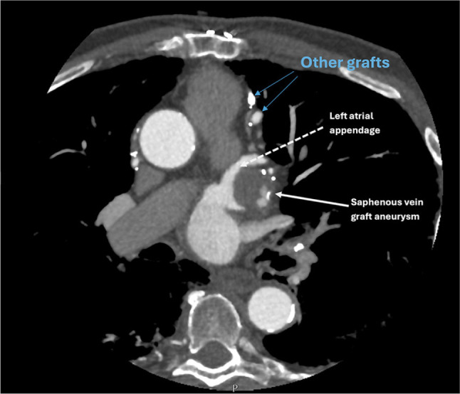
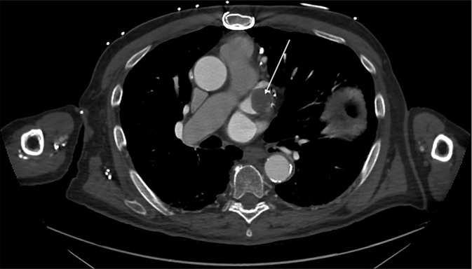
Saphenous vein graft aneurysm (SVGA) is a rare but potentially life-threatening complication of coronary artery bypass grafting (CABG). Its incidence is likely underreported due to asymptomatic cases and undiagnosed acute events. While SVGAs are more commonly associated with right atrial compression, presentation as a left atrial mass is rare. We present the case of an 85-year-old man with a history of CABG, who was incidentally found to have a left atrial appendage (LAA) density on a computed tomography (CT) chest, abdomen, and pelvis performed for unrelated symptoms of back pain and constipation. The density was initially suspected to be an LAA thrombus. However, a dedicated cardiac CT with delayed-phase imaging revealed a largely thrombosed aneurysmal saphenous vein graft to the obtuse marginal artery, which indented the LAA, mimicking an intracardiac mass. This case underscores the critical role of multimodality imaging, particularly cardiac CT, in differentiating vascular aneurysms from true intracardiac masses. Given the patient's asymptomatic status, conservative management with close follow-up was pursued. This case adds to the limited literature on SVGAs mimicking left atrial pathology and highlights the importance of recognizing this rare entity to avoid unnecessary interventions. It also emphasizes the evolving role of cardiac CT as a noninvasive, high-yield diagnostic tool for complex post-CABG anatomical assessments.
期刊介绍:
The Journal of Clinical Imaging Science (JCIS) is an open access peer-reviewed journal committed to publishing high-quality articles in the field of Imaging Science. The journal aims to present Imaging Science and relevant clinical information in an understandable and useful format. The journal is owned and published by the Scientific Scholar. Audience Our audience includes Radiologists, Researchers, Clinicians, medical professionals and students. Review process JCIS has a highly rigorous peer-review process that makes sure that manuscripts are scientifically accurate, relevant, novel and important. Authors disclose all conflicts, affiliations and financial associations such that the published content is not biased.


 求助内容:
求助内容: 应助结果提醒方式:


