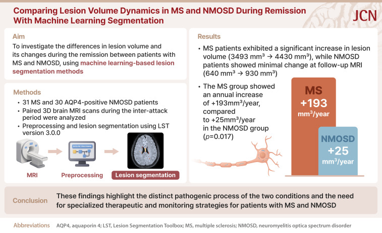
Comparing Lesion Volume Dynamics Between Multiple Sclerosis and Neuromyelitis Optica Spectrum Disorder During Remission Using Machine-Learning Segmentation.
Shaun G Hong, Ki Hoon Kim, You-Ri Kang, Jae-Won Hyun, Su-Hyun Kim, Ho Jin Kim
{"title":"Comparing Lesion Volume Dynamics Between Multiple Sclerosis and Neuromyelitis Optica Spectrum Disorder During Remission Using Machine-Learning Segmentation.","authors":"Shaun G Hong, Ki Hoon Kim, You-Ri Kang, Jae-Won Hyun, Su-Hyun Kim, Ho Jin Kim","doi":"10.3988/jcn.2025.0199","DOIUrl":null,"url":null,"abstract":"<p><strong>Background and purpose: </strong>Multiple sclerosis (MS) and neuromyelitis optica spectrum disorder (NMOSD) are inflammatory demyelinating conditions of the central nervous system that have distinct pathological mechanisms. There is a paucity of studies comparing the accumulation of subclinical lesions between MS and NMOSD, especially during the clinical remission period. Recent advances in neuroimaging techniques, those particularly involving the use of machine learning (ML) methods for lesion segmentation, have provided new opportunities to quantitatively assess the volumes of brain lesions and how they change over time. In this study, we aimed to use ML-based lesion segmentation methods to measure differences in lesion volumes and their changes during the remission period between patients with MS and NMOSD.</p><p><strong>Methods: </strong>This study included a retrospective cohort of 31 patients with MS and patients with 30 aquaporin-4-positive (AQP4⁺) NMOSD from the National Cancer Center registry. Serial 3D brain magnetic resonance imaging (MRI) scans obtained during the interattack period were analyzed using ML-based segmentation. MRI data preprocessing included alignment, distortion correction, and normalization, with lesion mapping and statistical analyses determining changes in lesion volumes.</p><p><strong>Results: </strong>The MS patients exhibited significant increases in the median lesion volume (from 3,493 mm³ to 4,430 mm³, <i>p</i><0.001), indicating ongoing subclinical activity without clinical relapses. In contrast, the NMOSD patients showed no significant change in the median lesion volume (from 640 mm³ to 930 mm³, <i>p</i>=0.129), supporting an attack-dependent disease course. The lesion volume increased by 193 mm³/year in the MS group, compared with only 25 mm³/year in the NMOSD group (<i>p</i>=0.017).</p><p><strong>Conclusions: </strong>These findings highlight the distinct pathogenic processes of the two conditions and hence the need for specialized therapeutic and monitoring strategies for patients with MS and AQP4⁺ NMOSD.</p>","PeriodicalId":15432,"journal":{"name":"Journal of Clinical Neurology","volume":"21 5","pages":"433-438"},"PeriodicalIF":3.1000,"publicationDate":"2025-09-01","publicationTypes":"Journal Article","fieldsOfStudy":null,"isOpenAccess":false,"openAccessPdf":"https://www.ncbi.nlm.nih.gov/pmc/articles/PMC12411297/pdf/","citationCount":"0","resultStr":null,"platform":"Semanticscholar","paperid":null,"PeriodicalName":"Journal of Clinical Neurology","FirstCategoryId":"3","ListUrlMain":"https://doi.org/10.3988/jcn.2025.0199","RegionNum":3,"RegionCategory":"医学","ArticlePicture":[],"TitleCN":null,"AbstractTextCN":null,"PMCID":null,"EPubDate":"","PubModel":"","JCR":"Q2","JCRName":"CLINICAL NEUROLOGY","Score":null,"Total":0}
引用次数: 0
Abstract
Background and purpose: Multiple sclerosis (MS) and neuromyelitis optica spectrum disorder (NMOSD) are inflammatory demyelinating conditions of the central nervous system that have distinct pathological mechanisms. There is a paucity of studies comparing the accumulation of subclinical lesions between MS and NMOSD, especially during the clinical remission period. Recent advances in neuroimaging techniques, those particularly involving the use of machine learning (ML) methods for lesion segmentation, have provided new opportunities to quantitatively assess the volumes of brain lesions and how they change over time. In this study, we aimed to use ML-based lesion segmentation methods to measure differences in lesion volumes and their changes during the remission period between patients with MS and NMOSD.
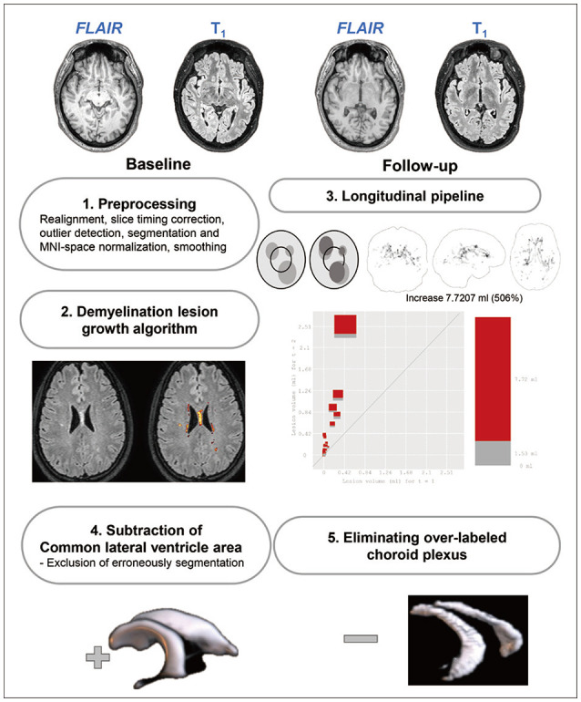
Methods: This study included a retrospective cohort of 31 patients with MS and patients with 30 aquaporin-4-positive (AQP4⁺) NMOSD from the National Cancer Center registry. Serial 3D brain magnetic resonance imaging (MRI) scans obtained during the interattack period were analyzed using ML-based segmentation. MRI data preprocessing included alignment, distortion correction, and normalization, with lesion mapping and statistical analyses determining changes in lesion volumes.
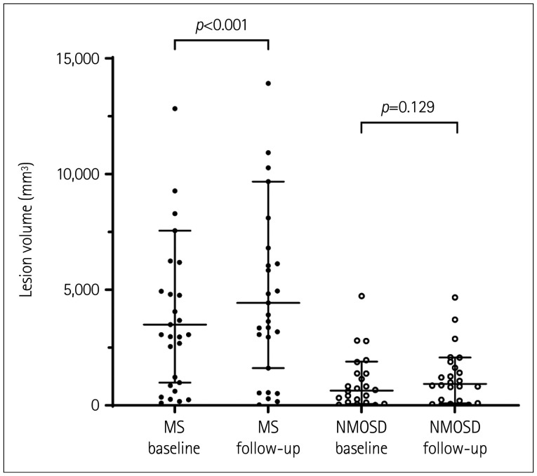
Results: The MS patients exhibited significant increases in the median lesion volume (from 3,493 mm³ to 4,430 mm³, p<0.001), indicating ongoing subclinical activity without clinical relapses. In contrast, the NMOSD patients showed no significant change in the median lesion volume (from 640 mm³ to 930 mm³, p=0.129), supporting an attack-dependent disease course. The lesion volume increased by 193 mm³/year in the MS group, compared with only 25 mm³/year in the NMOSD group (p=0.017).
Conclusions: These findings highlight the distinct pathogenic processes of the two conditions and hence the need for specialized therapeutic and monitoring strategies for patients with MS and AQP4⁺ NMOSD.
期刊介绍:
The JCN aims to publish the cutting-edge research from around the world. The JCN covers clinical and translational research for physicians and researchers in the field of neurology. Encompassing the entire neurological diseases, our main focus is on the common disorders including stroke, epilepsy, Parkinson''s disease, dementia, multiple sclerosis, headache, and peripheral neuropathy. Any authors affiliated with an accredited biomedical institution may submit manuscripts of original articles, review articles, and letters to the editor. The JCN will allow clinical neurologists to enrich their knowledge of patient management, education, and clinical or experimental research, and hence their professionalism.
 求助内容:
求助内容: 应助结果提醒方式:


