{"title":"Primary cardiac angiosarcoma - a diagnostic roller-coaster till fatality.","authors":"Bhupendra Kumar Sihag, Ajay Bahl, Sarthak Wadhera, Arnav Aggarwal, Mohsin Raj Mantoo, Atit A Gawalkar","doi":"10.34172/jcvtr.025.33285","DOIUrl":null,"url":null,"abstract":"<p><p>A 28-year-old male with a relatively short history of progressive dyspnea and a large pericardial effusion with tamponade was found to have an intracardiac mass localized in right atrium (RA) on echocardiography. Multimodality imaging revealed an irregular mass abutting the lateral wall of RA, with infiltration into surrounding pericardium and superior venacava. Positron emission tomography (PET) scan confirmed the mass as metabolically active lesion, along with uptake in mediastinal structures and lymph nodes. After an unrewarding percutaneous endomyocardial biopsy, open surgical biopsy was performed. Histologic examination confirmed the diagnosis of cardiac angiosarcoma. Unfortunately, patient had refractory shock and recurrent massive pericardial effusion (hemorrhagic) after biopsy and succumbed. The case highlights diagnostic dilemma of pericardial effusion in tuberculosis-endemic areas, role of multi-modality imaging in confirming cardiac malignancy and poor outcome of such patients.</p>","PeriodicalId":15207,"journal":{"name":"Journal of Cardiovascular and Thoracic Research","volume":"17 2","pages":"139-142"},"PeriodicalIF":0.7000,"publicationDate":"2025-06-28","publicationTypes":"Journal Article","fieldsOfStudy":null,"isOpenAccess":false,"openAccessPdf":"https://www.ncbi.nlm.nih.gov/pmc/articles/PMC12375422/pdf/","citationCount":"0","resultStr":null,"platform":"Semanticscholar","paperid":null,"PeriodicalName":"Journal of Cardiovascular and Thoracic Research","FirstCategoryId":"1085","ListUrlMain":"https://doi.org/10.34172/jcvtr.025.33285","RegionNum":0,"RegionCategory":null,"ArticlePicture":[],"TitleCN":null,"AbstractTextCN":null,"PMCID":null,"EPubDate":"2025/6/1 0:00:00","PubModel":"eCollection","JCR":"Q3","JCRName":"CARDIAC & CARDIOVASCULAR SYSTEMS","Score":null,"Total":0}
引用次数: 0
Abstract
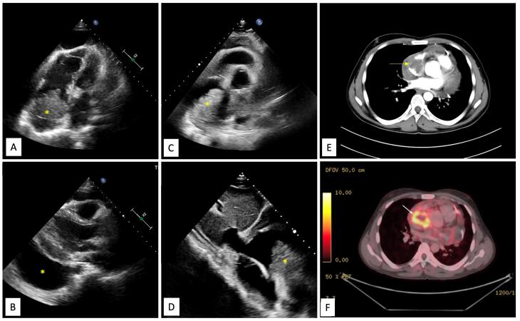
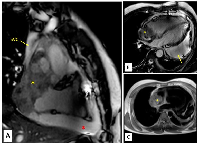
A 28-year-old male with a relatively short history of progressive dyspnea and a large pericardial effusion with tamponade was found to have an intracardiac mass localized in right atrium (RA) on echocardiography. Multimodality imaging revealed an irregular mass abutting the lateral wall of RA, with infiltration into surrounding pericardium and superior venacava. Positron emission tomography (PET) scan confirmed the mass as metabolically active lesion, along with uptake in mediastinal structures and lymph nodes. After an unrewarding percutaneous endomyocardial biopsy, open surgical biopsy was performed. Histologic examination confirmed the diagnosis of cardiac angiosarcoma. Unfortunately, patient had refractory shock and recurrent massive pericardial effusion (hemorrhagic) after biopsy and succumbed. The case highlights diagnostic dilemma of pericardial effusion in tuberculosis-endemic areas, role of multi-modality imaging in confirming cardiac malignancy and poor outcome of such patients.

 求助内容:
求助内容: 应助结果提醒方式:


