Seoyeon Park, Soo Jin Cho, Sung Mok Kim, Moon Young Kim, Yeon Hyeon Choe
{"title":"Late Gadolinium Enhancement Variation in Asymptomatic Individuals: Comparison with Dilated Cardiomyopathy.","authors":"Seoyeon Park, Soo Jin Cho, Sung Mok Kim, Moon Young Kim, Yeon Hyeon Choe","doi":"10.3390/jcdd12080312","DOIUrl":null,"url":null,"abstract":"<p><p>Late gadolinium enhancements (LGEs) appear in asymptomatic individuals as septal stripes, which mimic abnormal LGEs in patients with dilated cardiomyopathy (DCM). We aimed to evaluate the frequency and extent of LGE variation in asymptomatic individuals and to compare it with those of DCM group. This retrospective study included asymptomatic and DCM groups who underwent CMR imaging. LGE was defined as a myocardial signal intensity higher than five standard-deviations of normal myocardium. LGE was evaluated in right ventricular insertion points (RVIPs) and mid-interventricular septum. A total of 273 asymptomatic individuals (age, 54.3 ± 5.8 years, 209 males) and 100 patients with DCM (age, 55.3 ± 4.9 years, 73 males) were included. LGE was observed in 99.3% of asymptomatic and 100% of DCM groups. The average number of myocardial segments with LGE was distinguishable between asymptomatic and DCM groups (5.5 ± 1.7 vs. 7.6 ± 2.2; <i>p</i> < 0.001). The thickness of LGE differed between two groups in mid-septum (4.5 ± 1.3 mm vs. 5.7 ± 1.8 mm; <i>p</i> < 0.001), upper RVIP (6.1 ± 1.9 mm vs. 8.7 ± 2.7 mm; <i>p</i> < 0.001), and lower RVIP (6.4 ± 2.3 mm vs. 8.6 ± 2.8 mm; <i>p</i> < 0.001). Considerable overlap was observed in LGE between asymptomatic and DCM groups despite different LGE characteristics between them. LGEs within normal range should not be interpreted as abnormal findings in the evaluation of myocardial diseases including DCM.</p>","PeriodicalId":15197,"journal":{"name":"Journal of Cardiovascular Development and Disease","volume":"12 8","pages":""},"PeriodicalIF":2.3000,"publicationDate":"2025-08-18","publicationTypes":"Journal Article","fieldsOfStudy":null,"isOpenAccess":false,"openAccessPdf":"https://www.ncbi.nlm.nih.gov/pmc/articles/PMC12386344/pdf/","citationCount":"0","resultStr":null,"platform":"Semanticscholar","paperid":null,"PeriodicalName":"Journal of Cardiovascular Development and Disease","FirstCategoryId":"3","ListUrlMain":"https://doi.org/10.3390/jcdd12080312","RegionNum":4,"RegionCategory":"医学","ArticlePicture":[],"TitleCN":null,"AbstractTextCN":null,"PMCID":null,"EPubDate":"","PubModel":"","JCR":"Q2","JCRName":"CARDIAC & CARDIOVASCULAR SYSTEMS","Score":null,"Total":0}
引用次数: 0
Abstract
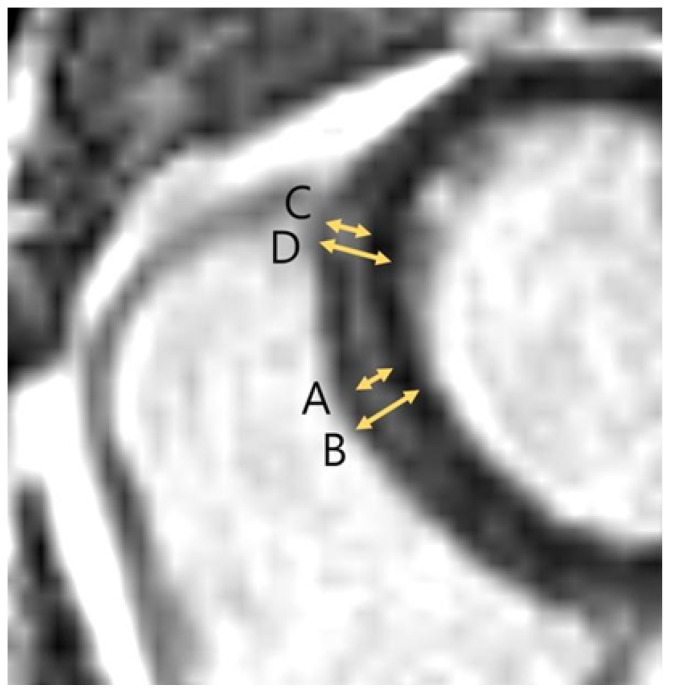
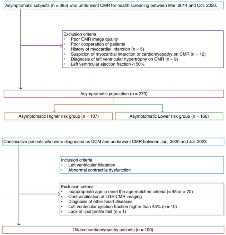
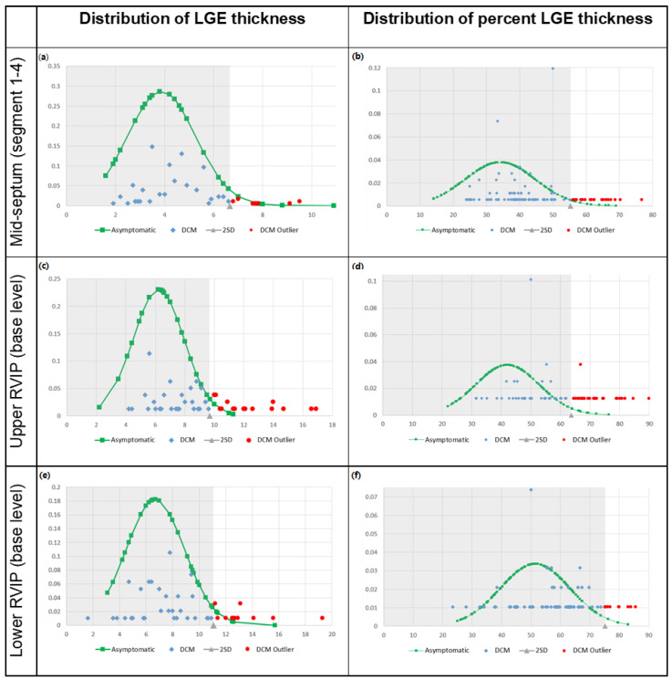
Late gadolinium enhancements (LGEs) appear in asymptomatic individuals as septal stripes, which mimic abnormal LGEs in patients with dilated cardiomyopathy (DCM). We aimed to evaluate the frequency and extent of LGE variation in asymptomatic individuals and to compare it with those of DCM group. This retrospective study included asymptomatic and DCM groups who underwent CMR imaging. LGE was defined as a myocardial signal intensity higher than five standard-deviations of normal myocardium. LGE was evaluated in right ventricular insertion points (RVIPs) and mid-interventricular septum. A total of 273 asymptomatic individuals (age, 54.3 ± 5.8 years, 209 males) and 100 patients with DCM (age, 55.3 ± 4.9 years, 73 males) were included. LGE was observed in 99.3% of asymptomatic and 100% of DCM groups. The average number of myocardial segments with LGE was distinguishable between asymptomatic and DCM groups (5.5 ± 1.7 vs. 7.6 ± 2.2; p < 0.001). The thickness of LGE differed between two groups in mid-septum (4.5 ± 1.3 mm vs. 5.7 ± 1.8 mm; p < 0.001), upper RVIP (6.1 ± 1.9 mm vs. 8.7 ± 2.7 mm; p < 0.001), and lower RVIP (6.4 ± 2.3 mm vs. 8.6 ± 2.8 mm; p < 0.001). Considerable overlap was observed in LGE between asymptomatic and DCM groups despite different LGE characteristics between them. LGEs within normal range should not be interpreted as abnormal findings in the evaluation of myocardial diseases including DCM.
晚期钆增强(LGEs)在无症状个体中表现为间隔条纹,类似于扩张型心肌病(DCM)患者的异常LGEs。我们的目的是评估无症状个体LGE变异的频率和程度,并将其与DCM组进行比较。这项回顾性研究包括无症状组和DCM组,他们接受了CMR成像。LGE定义为心肌信号强度高于正常心肌5个标准差。在右心室插入点(RVIPs)和中室间隔处评估LGE。共纳入无症状患者273例(年龄54.3±5.8岁,男性209例)和DCM患者100例(年龄55.3±4.9岁,男性73例)。无症状组的LGE发生率为99.3%,DCM组的LGE发生率为100%。无症状组和DCM组LGE的平均心肌节段数可区分(5.5±1.7 vs. 7.6±2.2;p < 0.001)。两组间LGE厚度在中隔(4.5±1.3 mm vs. 5.7±1.8 mm, p < 0.001)、RVIP上部(6.1±1.9 mm vs. 8.7±2.7 mm, p < 0.001)和RVIP下部(6.4±2.3 mm vs. 8.6±2.8 mm, p < 0.001)存在差异。尽管无症状组和DCM组之间的LGE特征不同,但在LGE中观察到相当大的重叠。正常范围内的lge不应被解释为评估包括DCM在内的心肌疾病的异常发现。

 求助内容:
求助内容: 应助结果提醒方式:


